Robust 3D Segmentation of Pulmonary Nodules in Multislice CT Images
Kazunori Okada, Dorin Comaniciu
Real-Time Vision and Modeling
Department, Siemens Corporate Research, Inc.
kazokada@sfsu.edu
dorin.comaniciu@siemens.com
Arun Krishnan
CAD Program, Siemens
Medical Solutions USA, Inc.
arun.krishnan@siemens.com
We propose a robust and accurate algorithm for segmenting the 3D pulmonary
nodules in multislice CT scans. The solution unifies i) the parametric
anisotropic Gaussian model fitting of the volumetric data evaluated in Gaussian
scale-space and ii) non-parametric 3D segmentation based on normalized gradient
(mean shift) ascent defining the basin of attraction of the target tumor in the
4D spatial-intensity joint space. This unification, by treating the parametric
estimation results from the first step as a normal prior, realizes an efficient
3D tumor segmentation according to both spatial and intensity proximities
simultaneously. Experimental results show that the system reliably segments a
variety of nodules including part- or non-solid nodules which poses difficulty
for the existing solutions. The system also efficiently processes a
32x32x32-voxel volume-of-interest by six seconds on average.
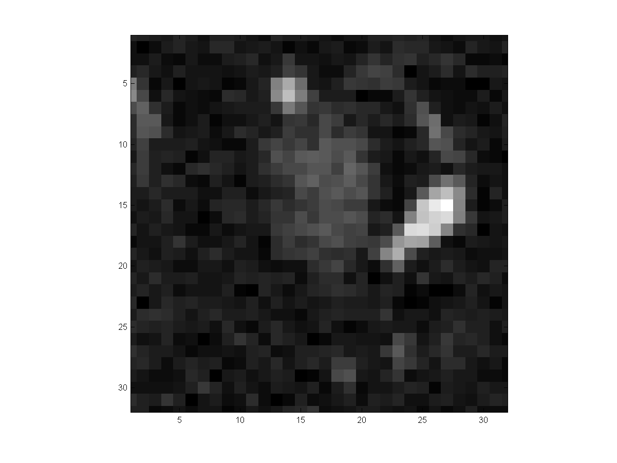
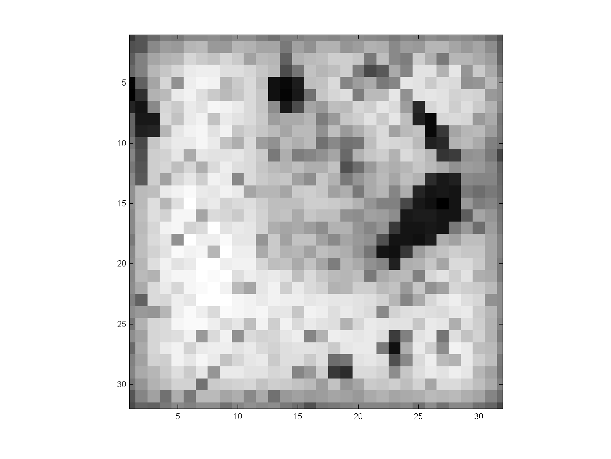
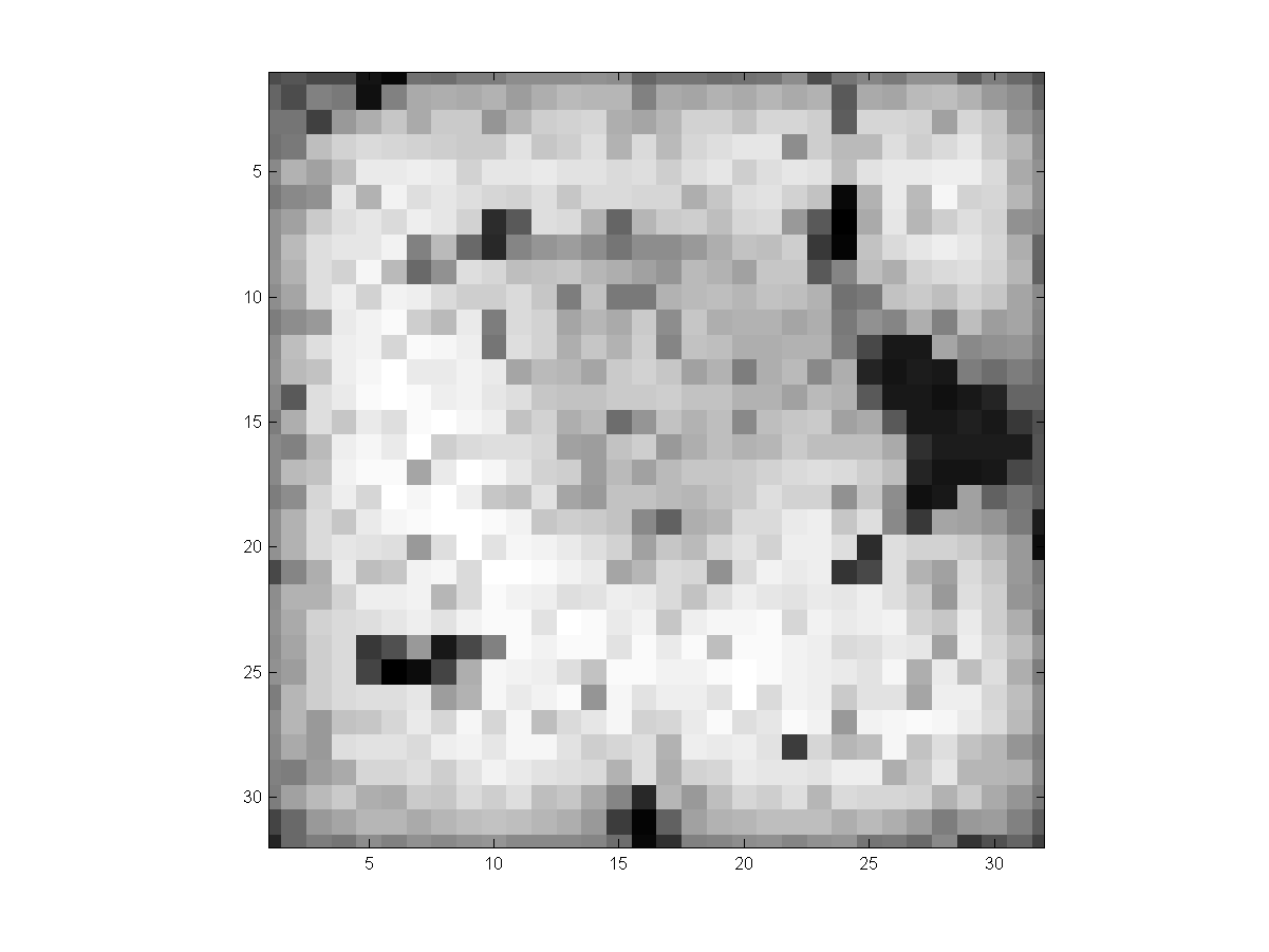
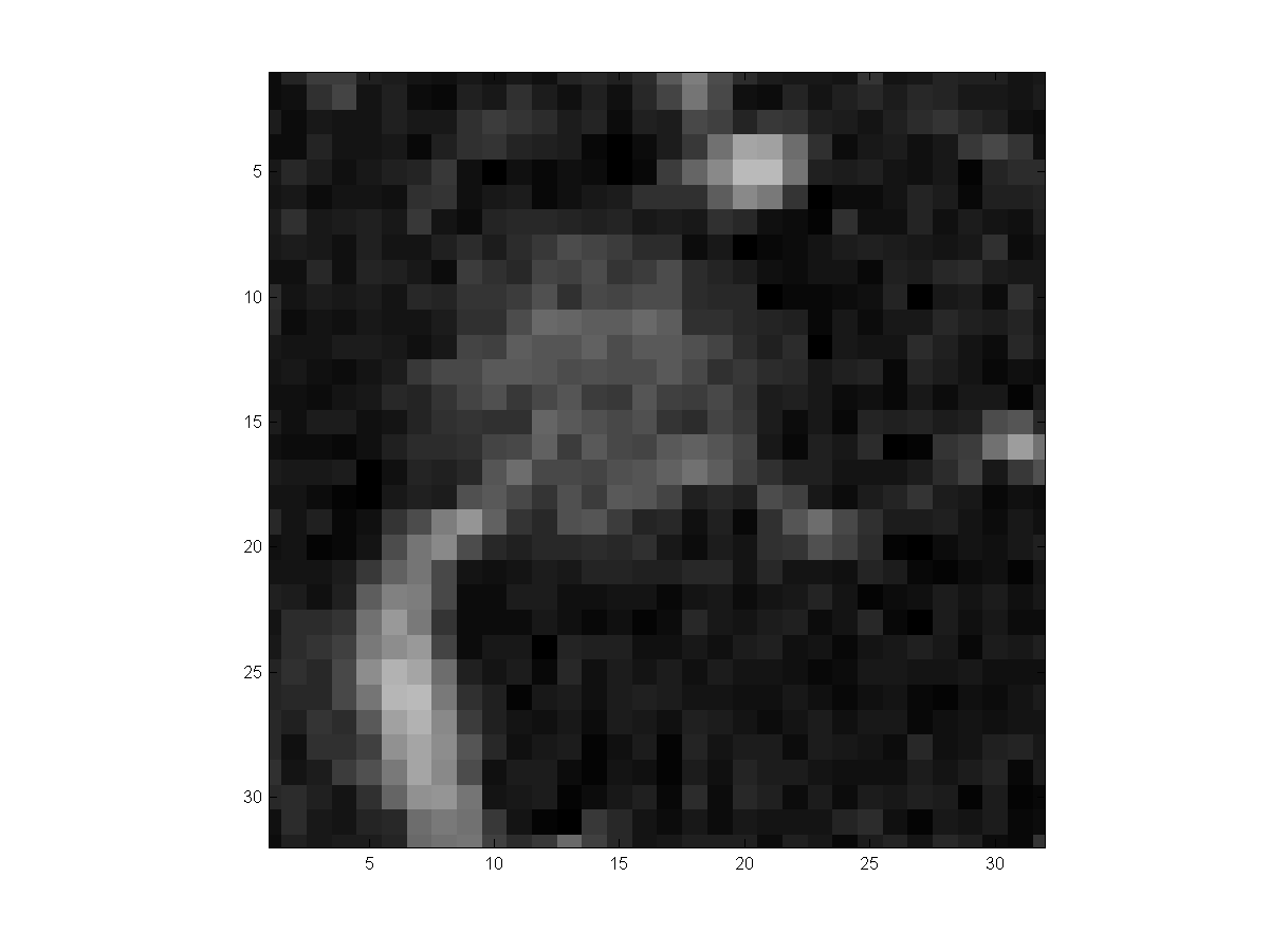
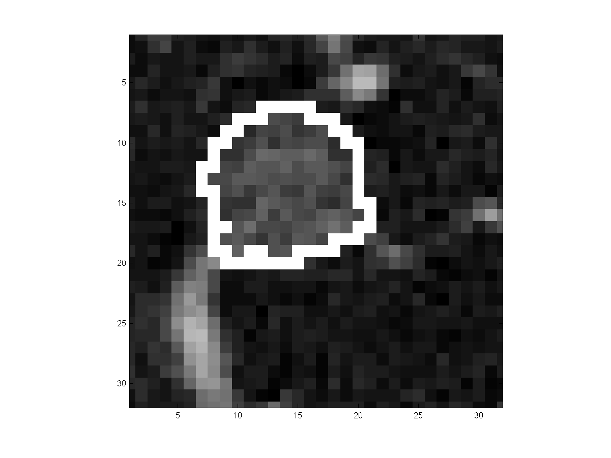
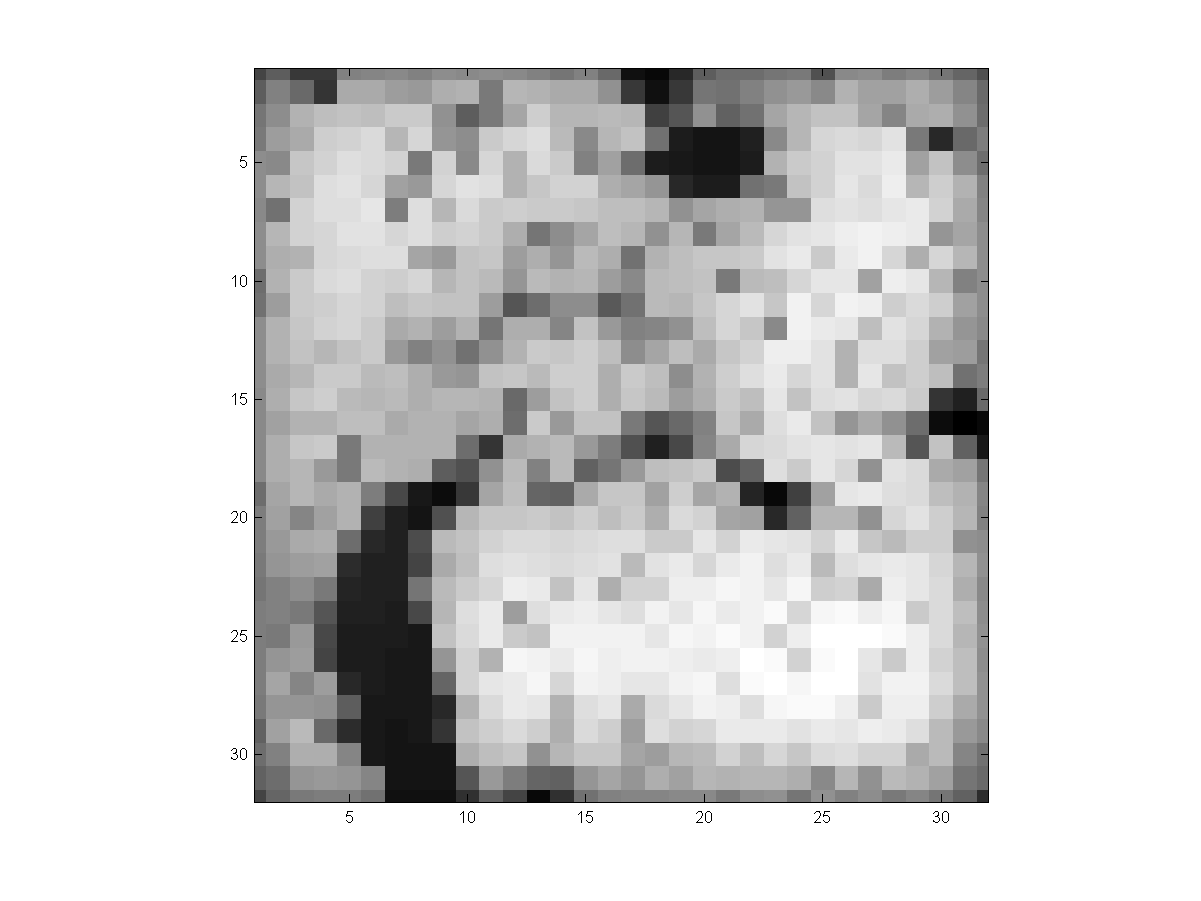
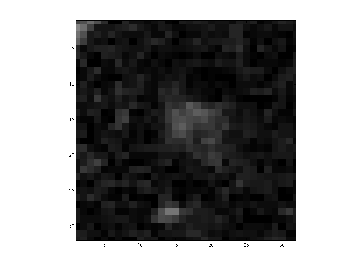
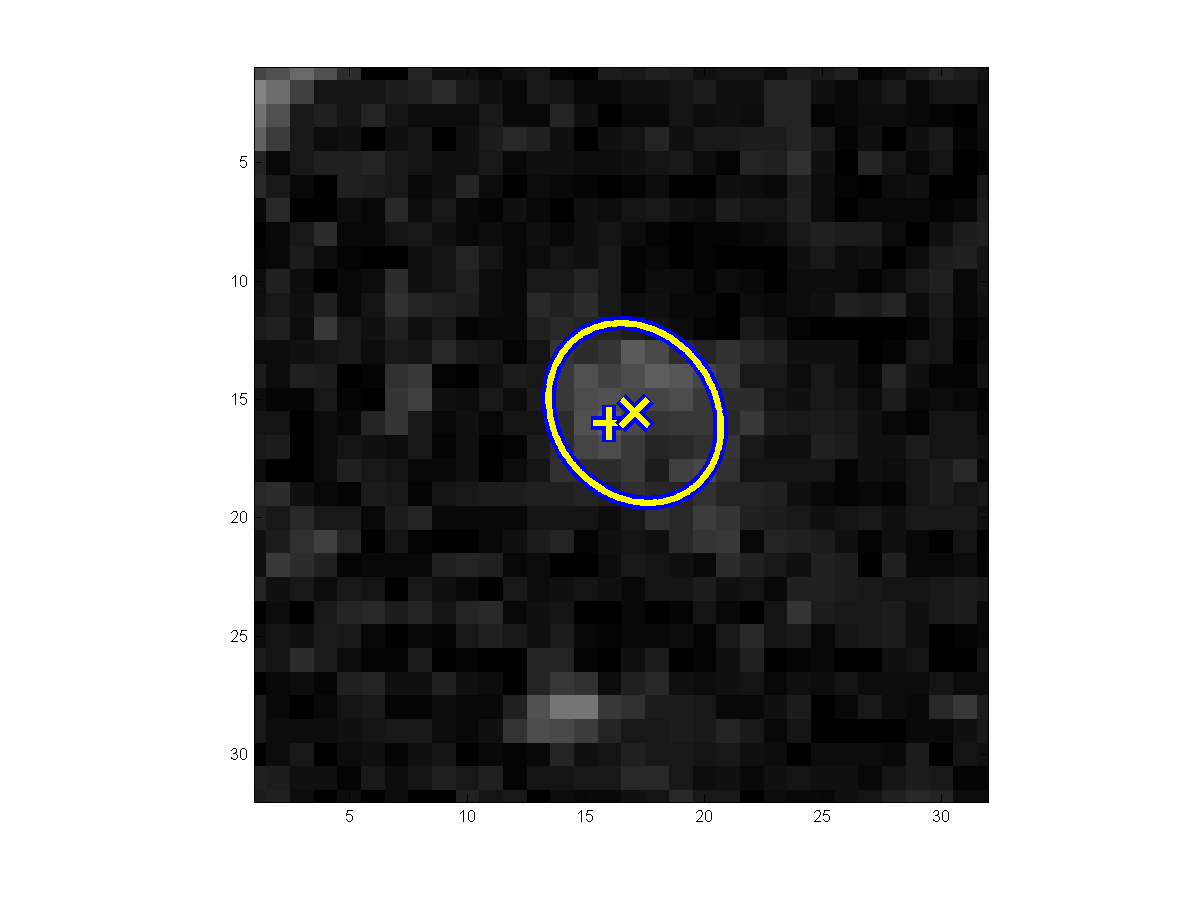
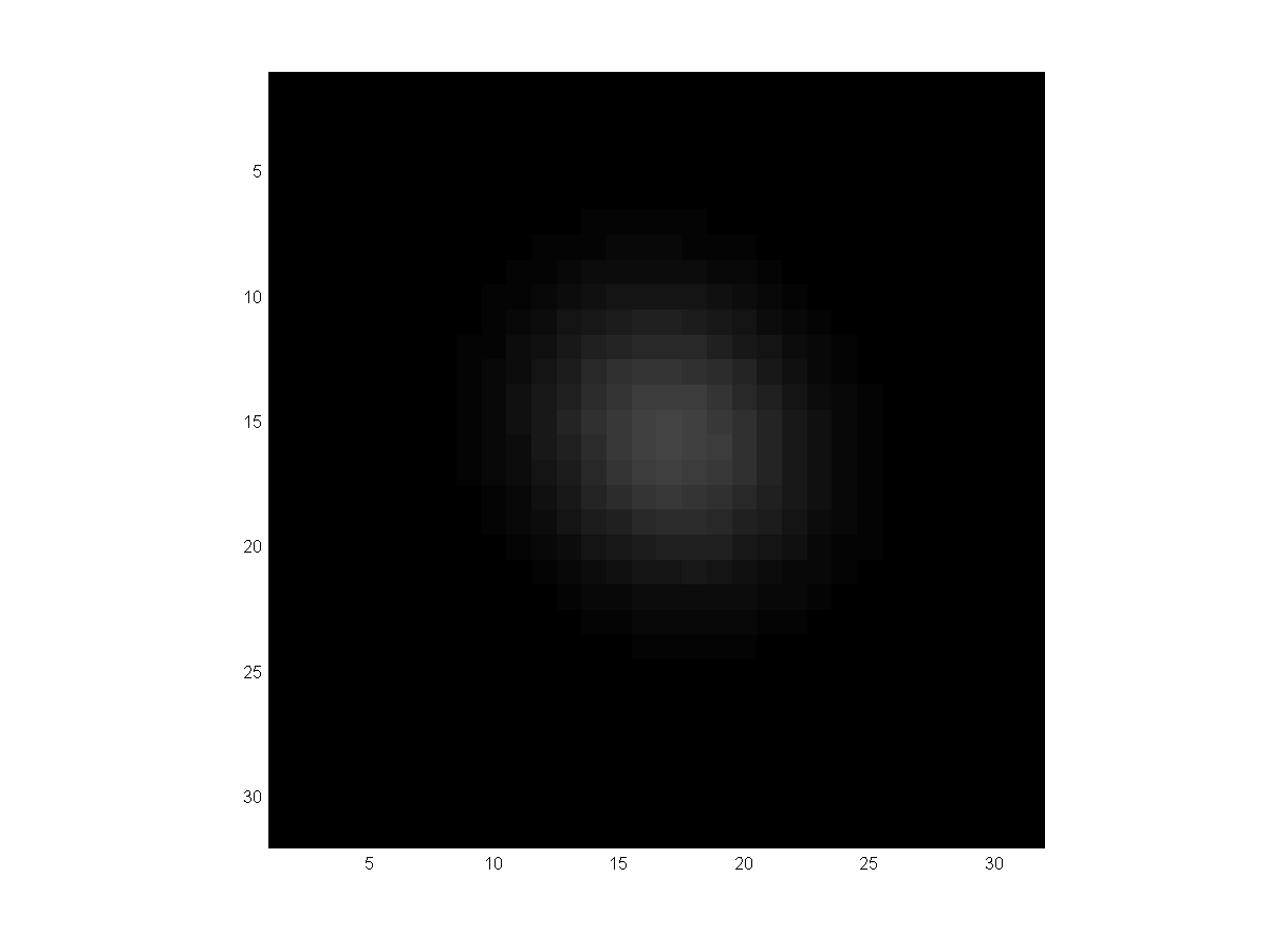
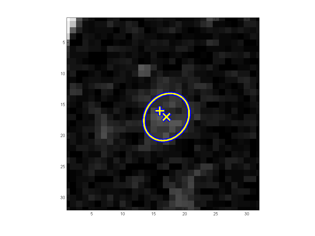
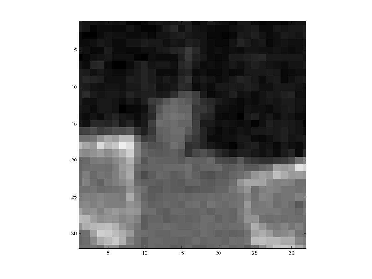
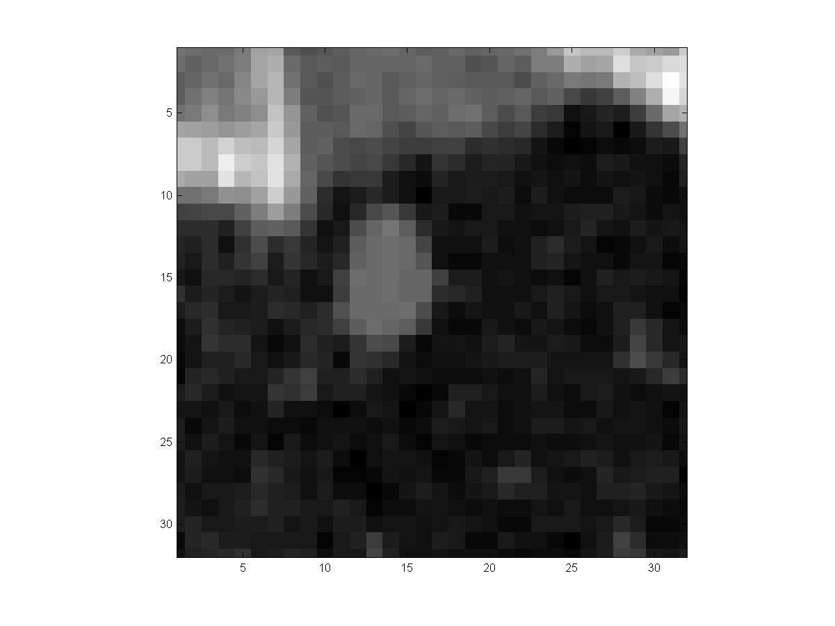
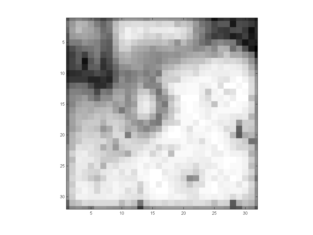
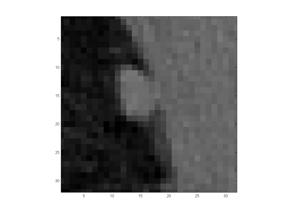
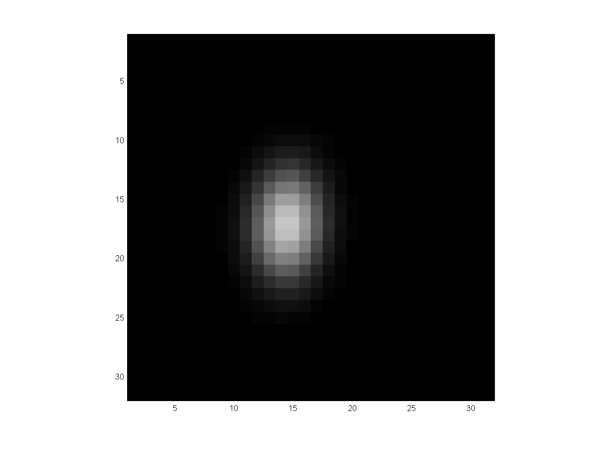
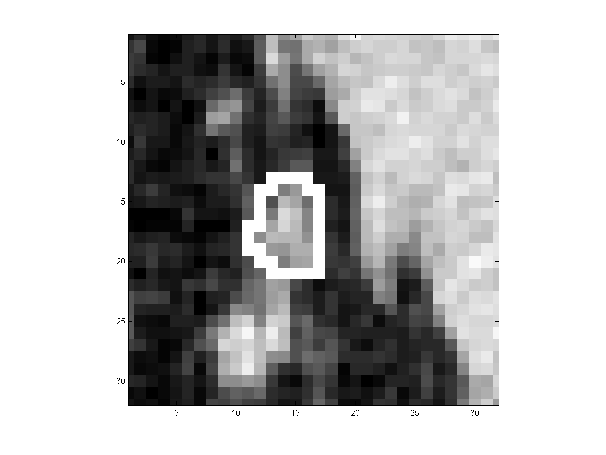
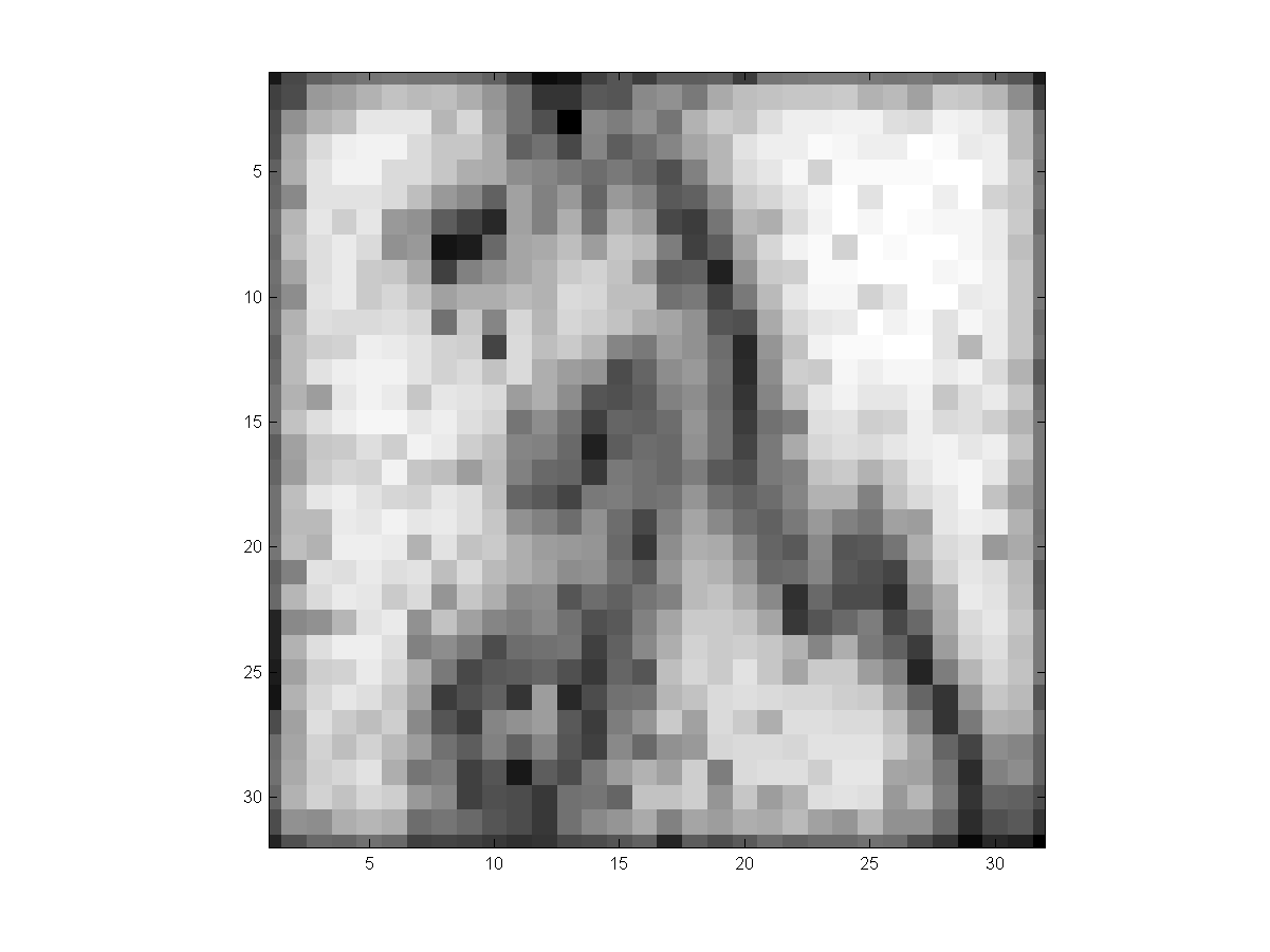
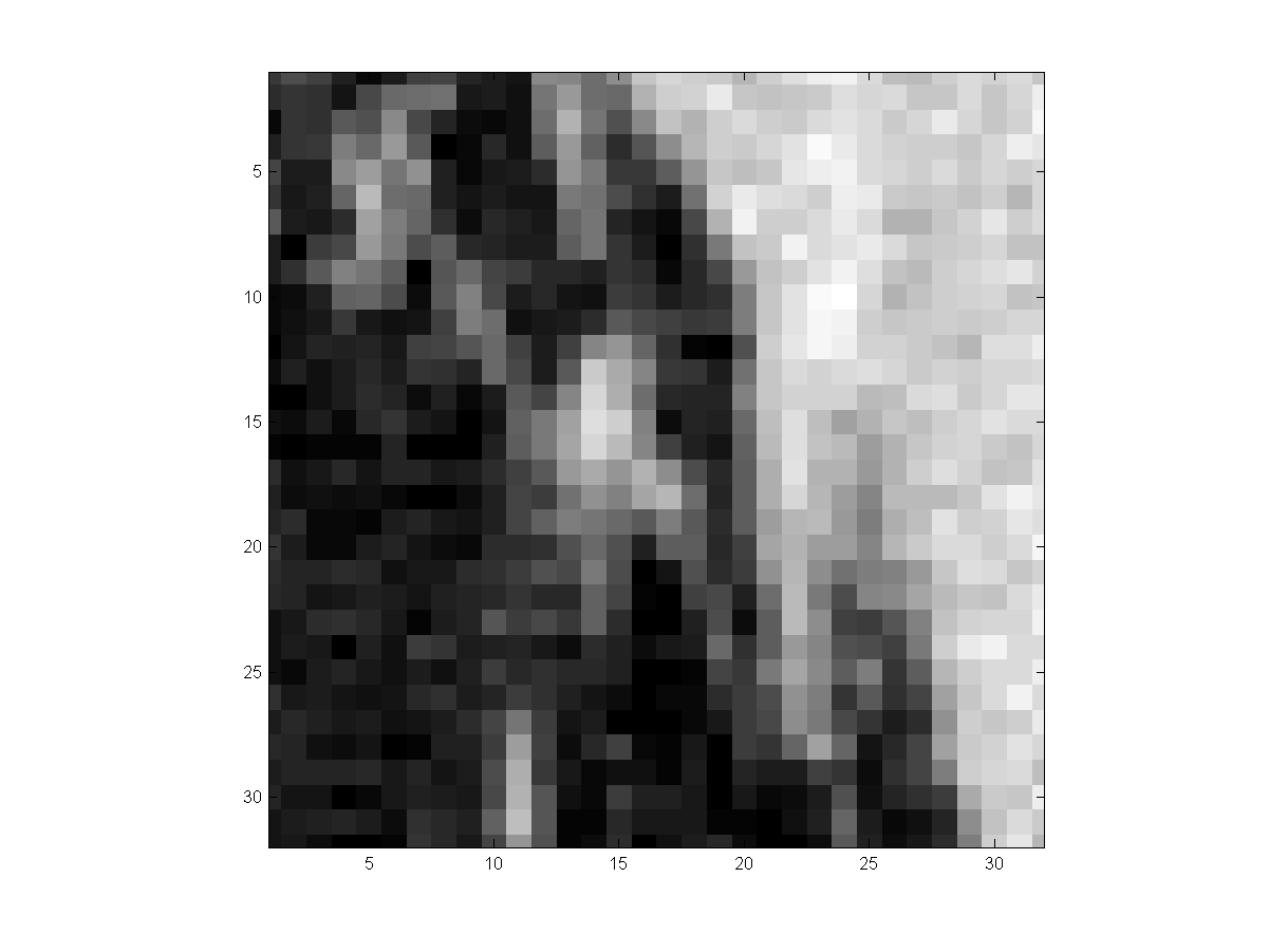
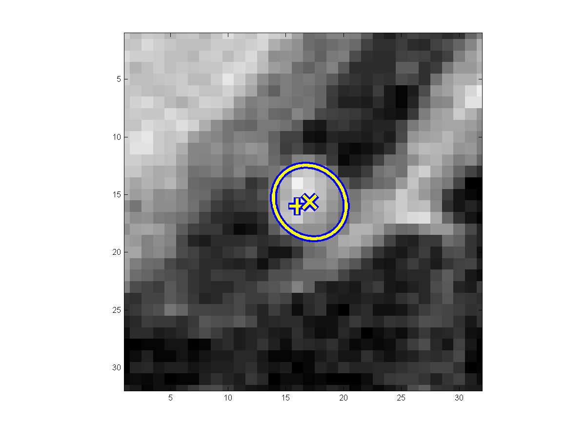
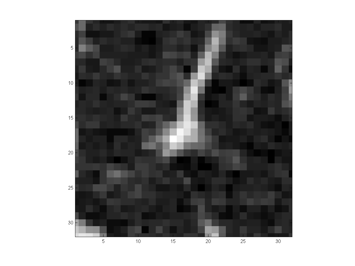
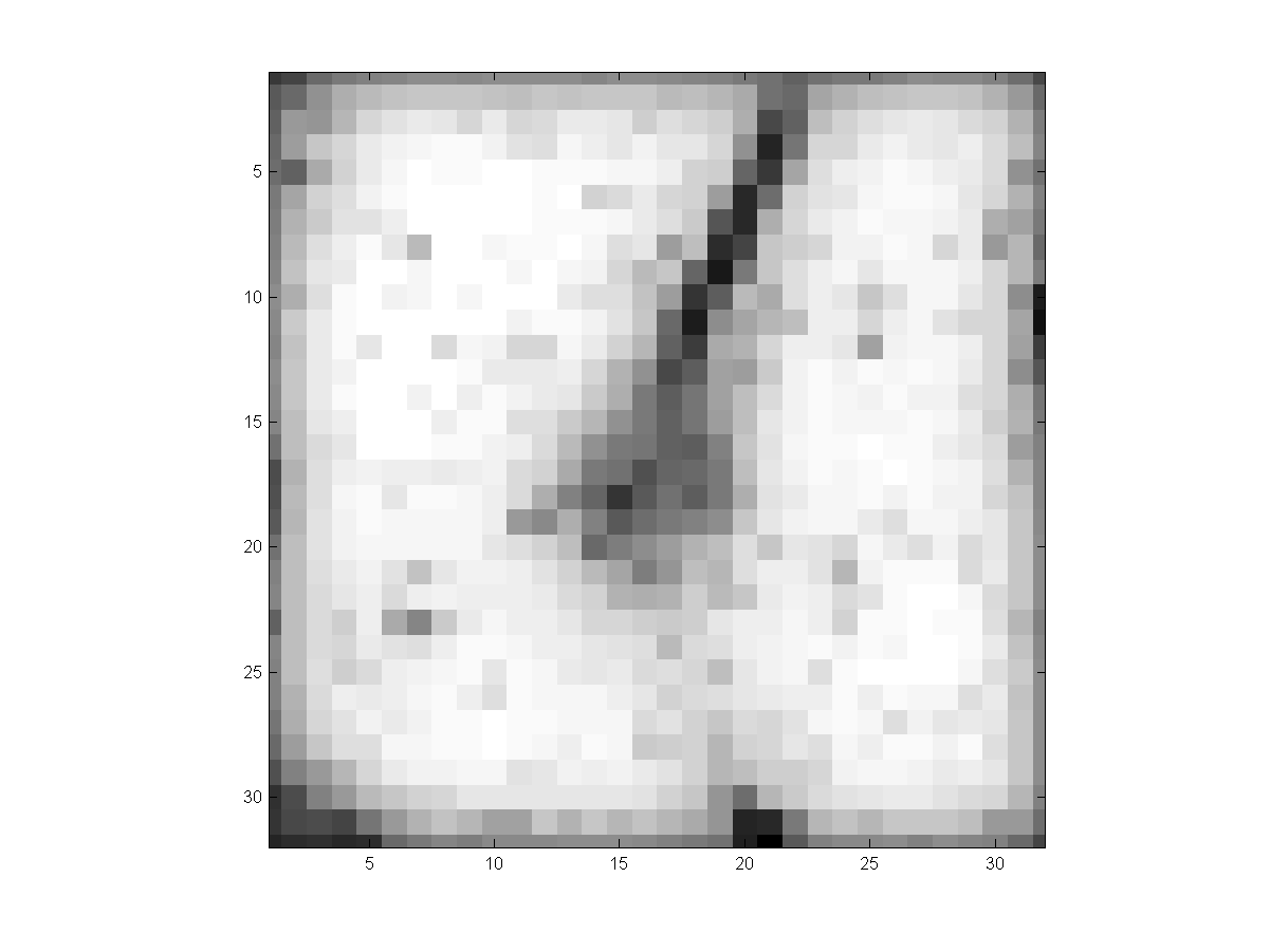
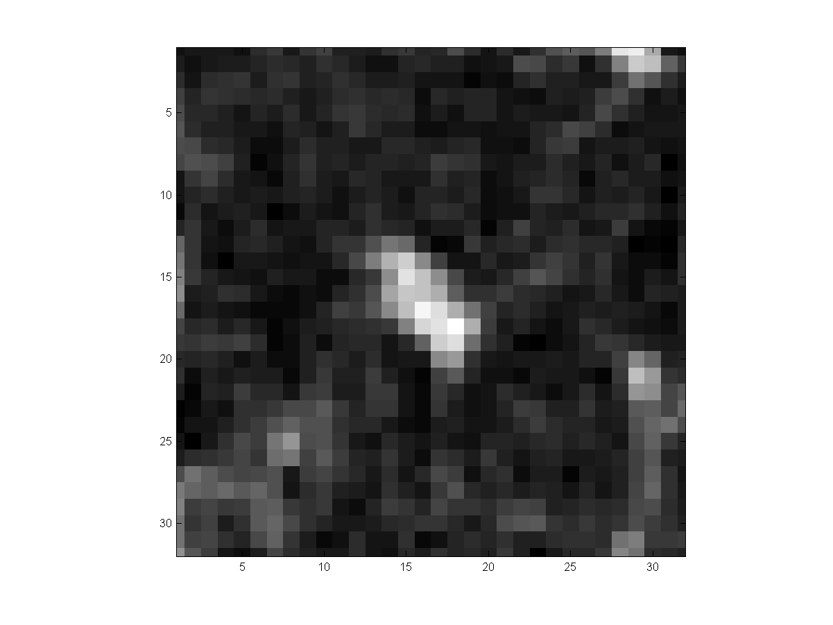
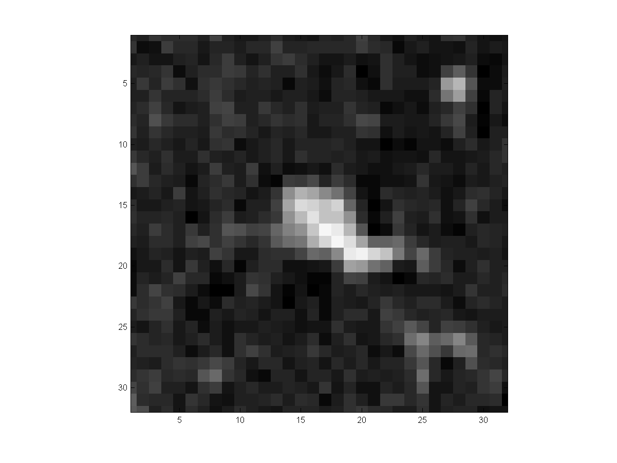
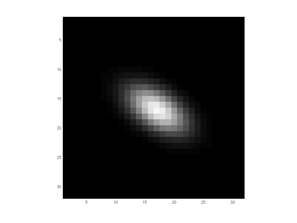
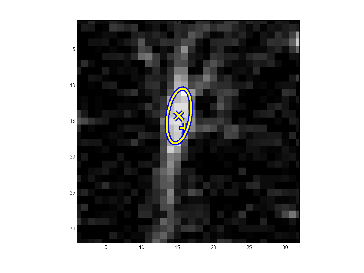
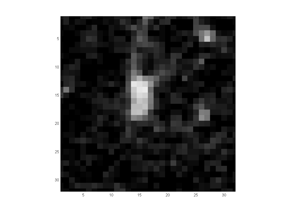
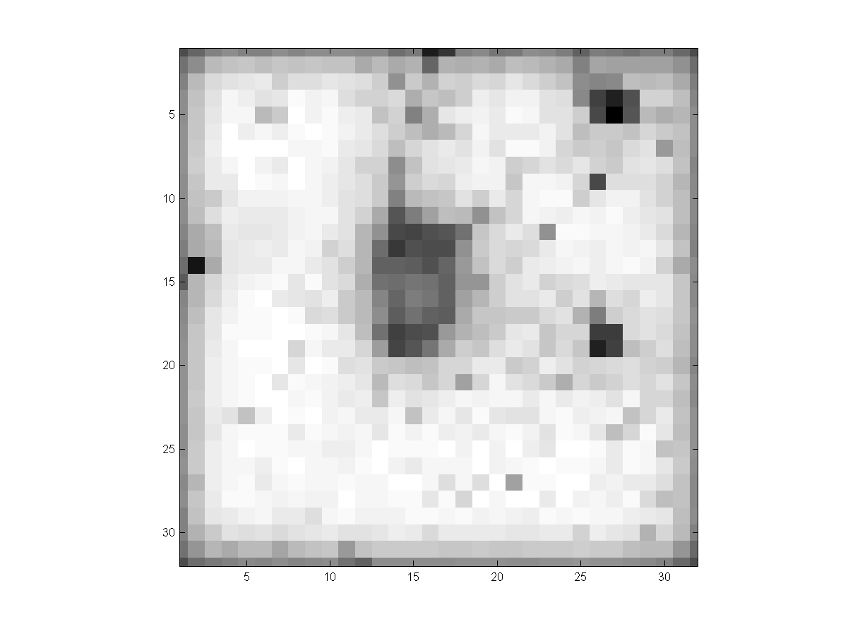
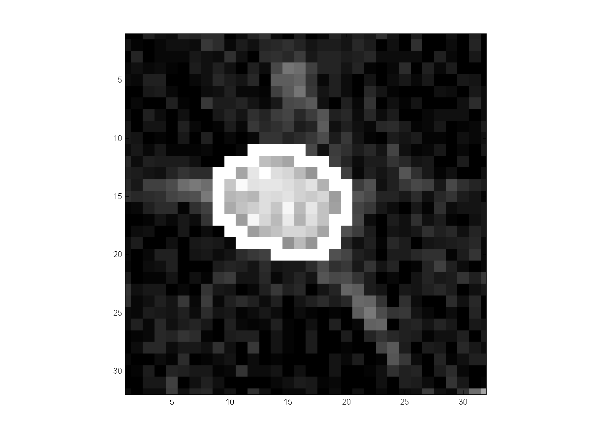
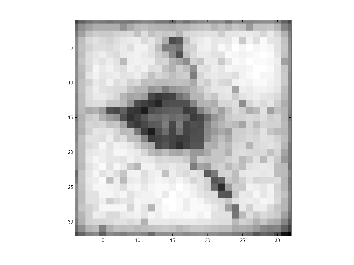
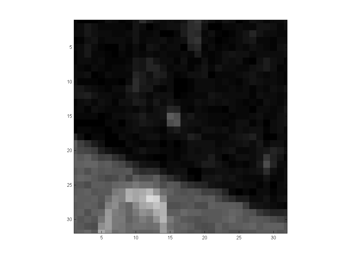
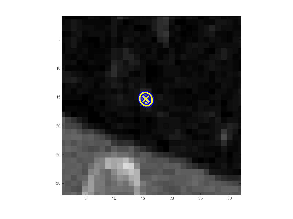
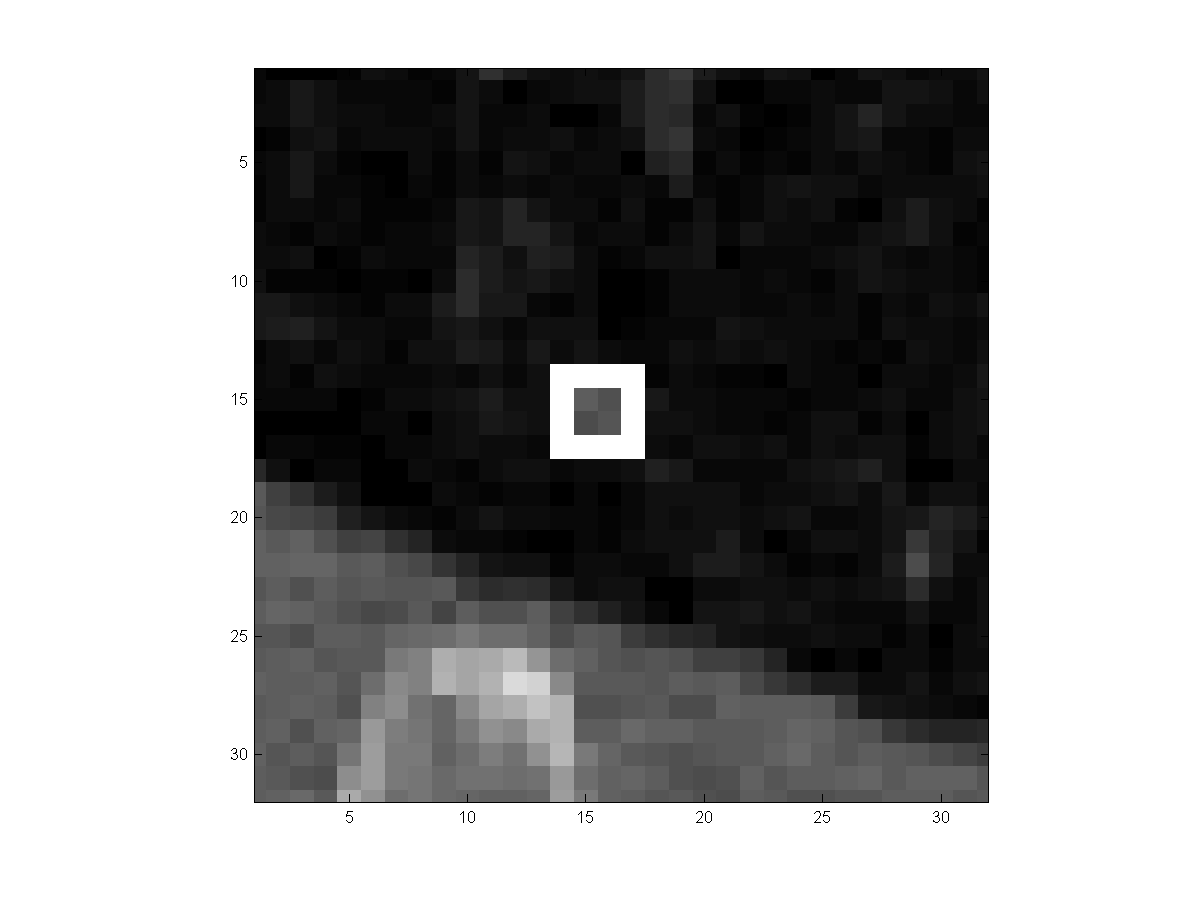
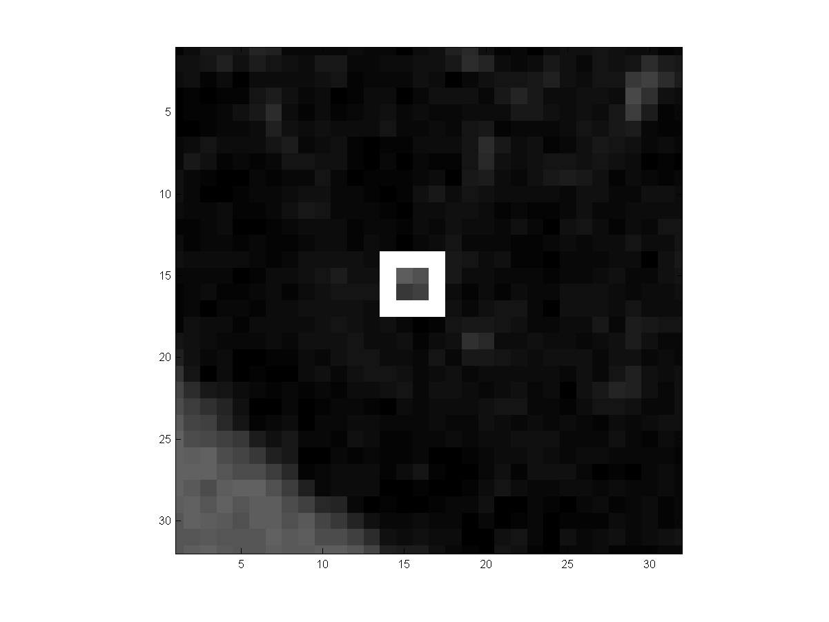
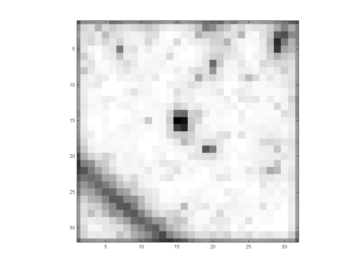
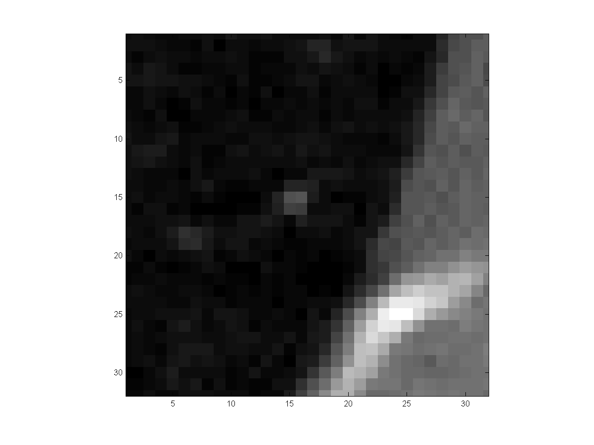
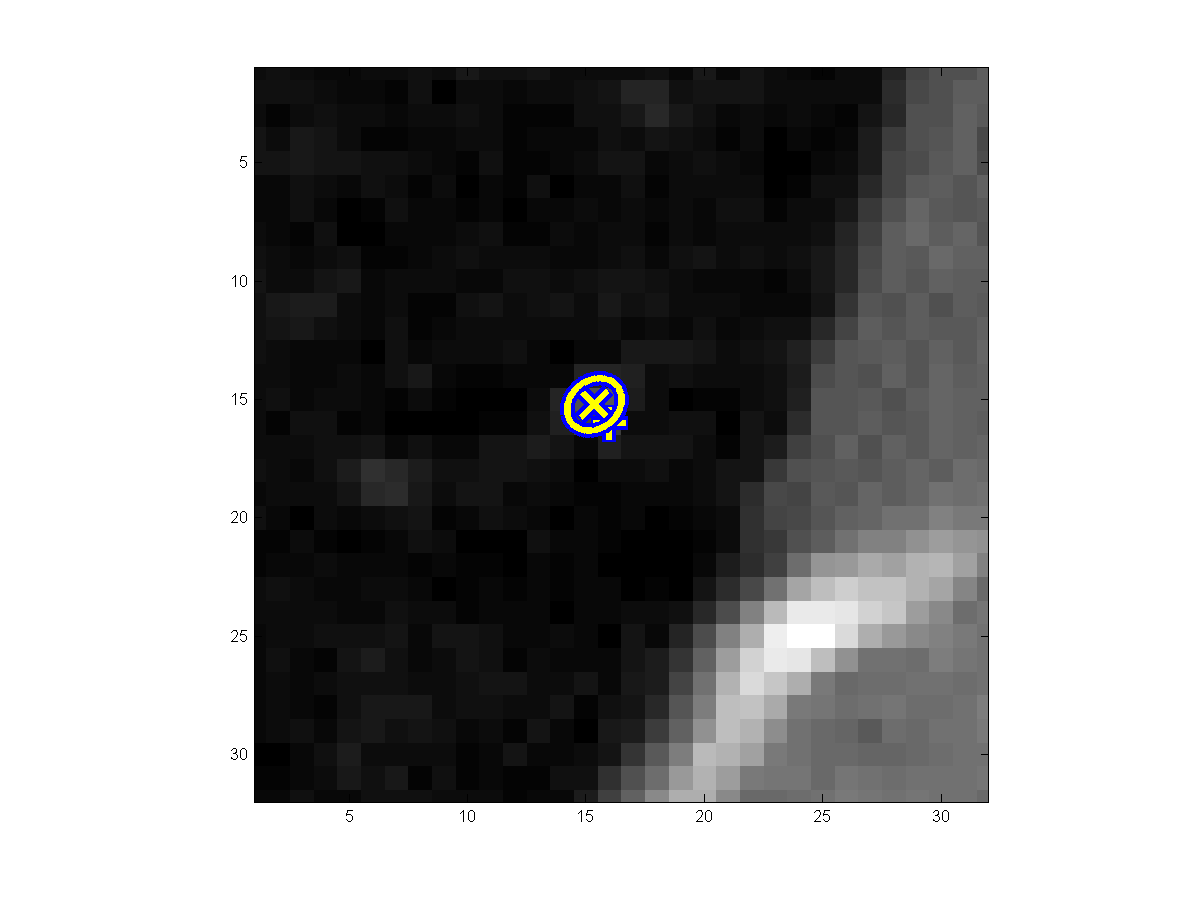
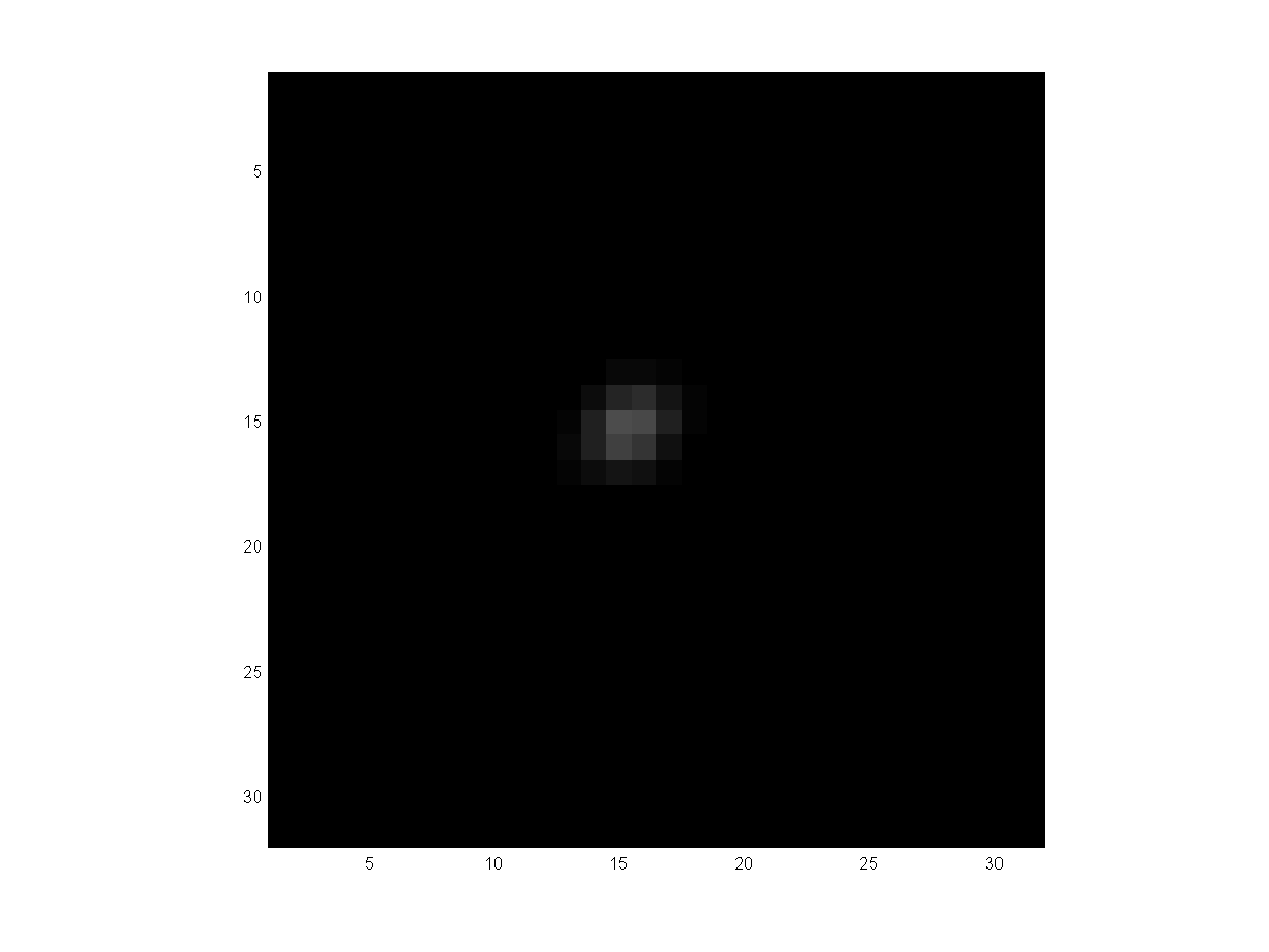
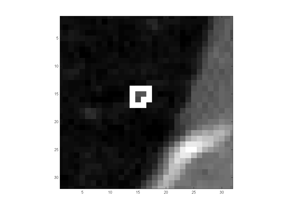
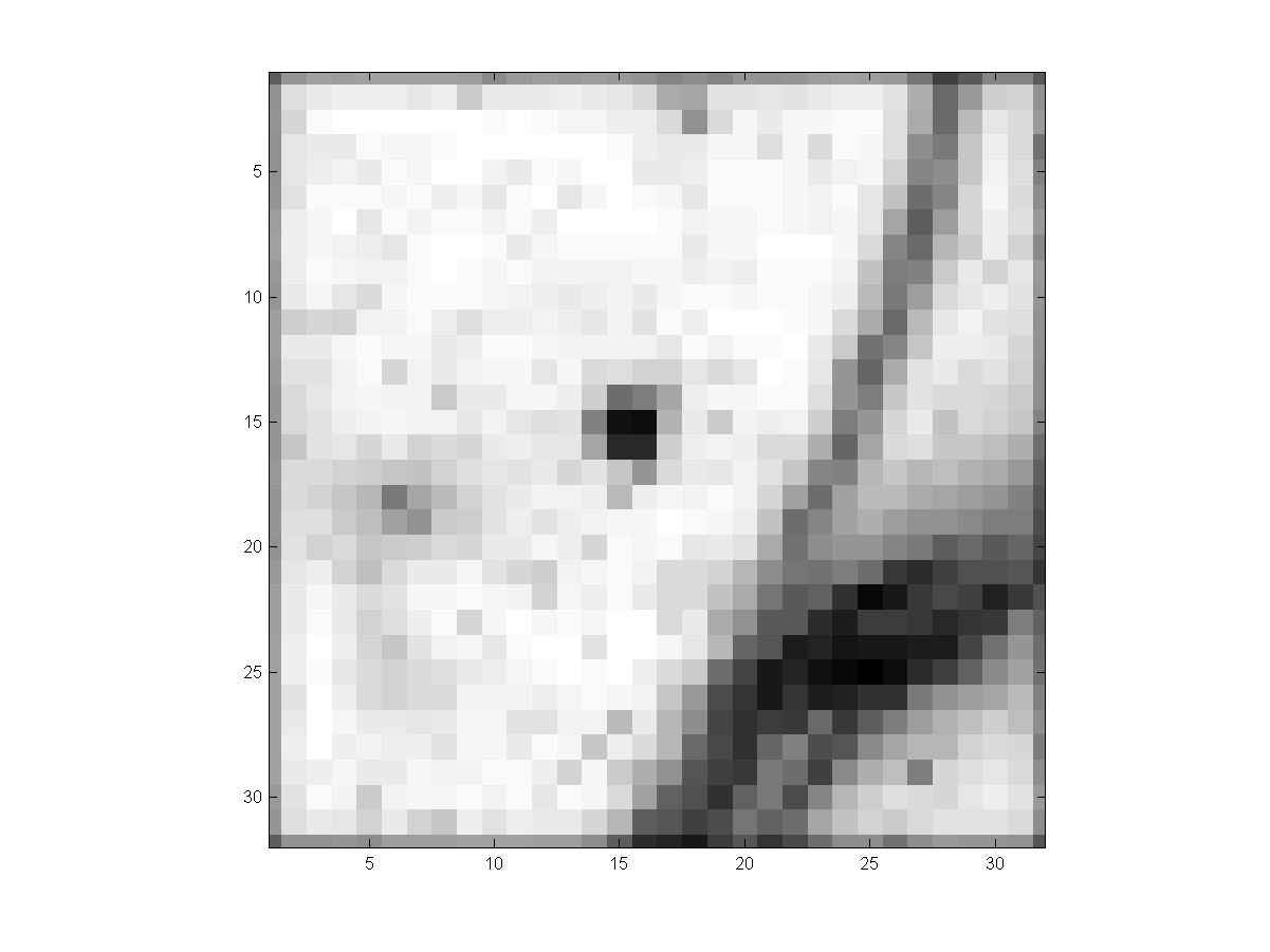
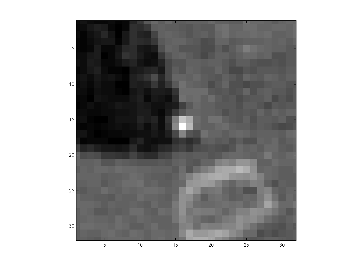
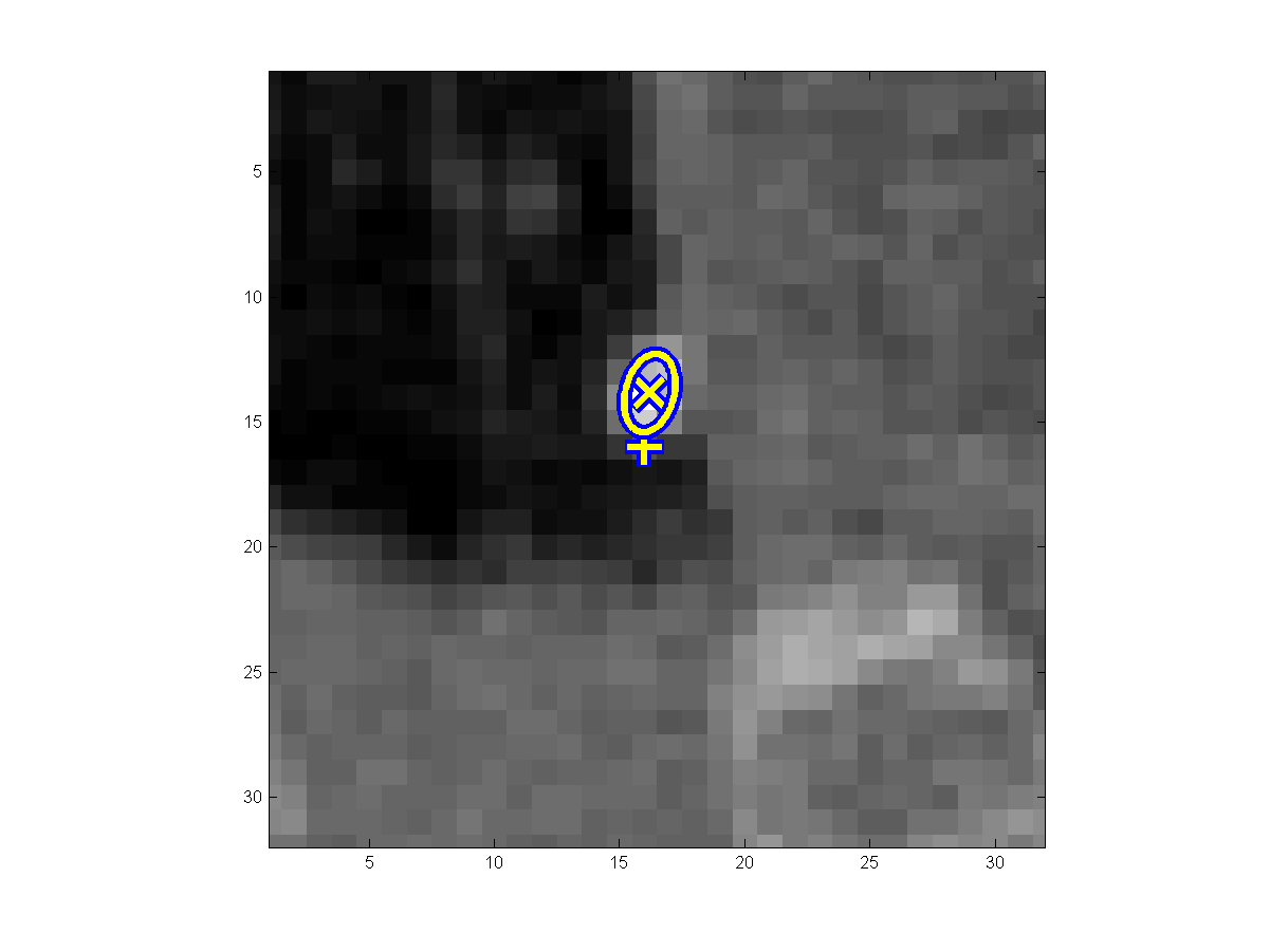
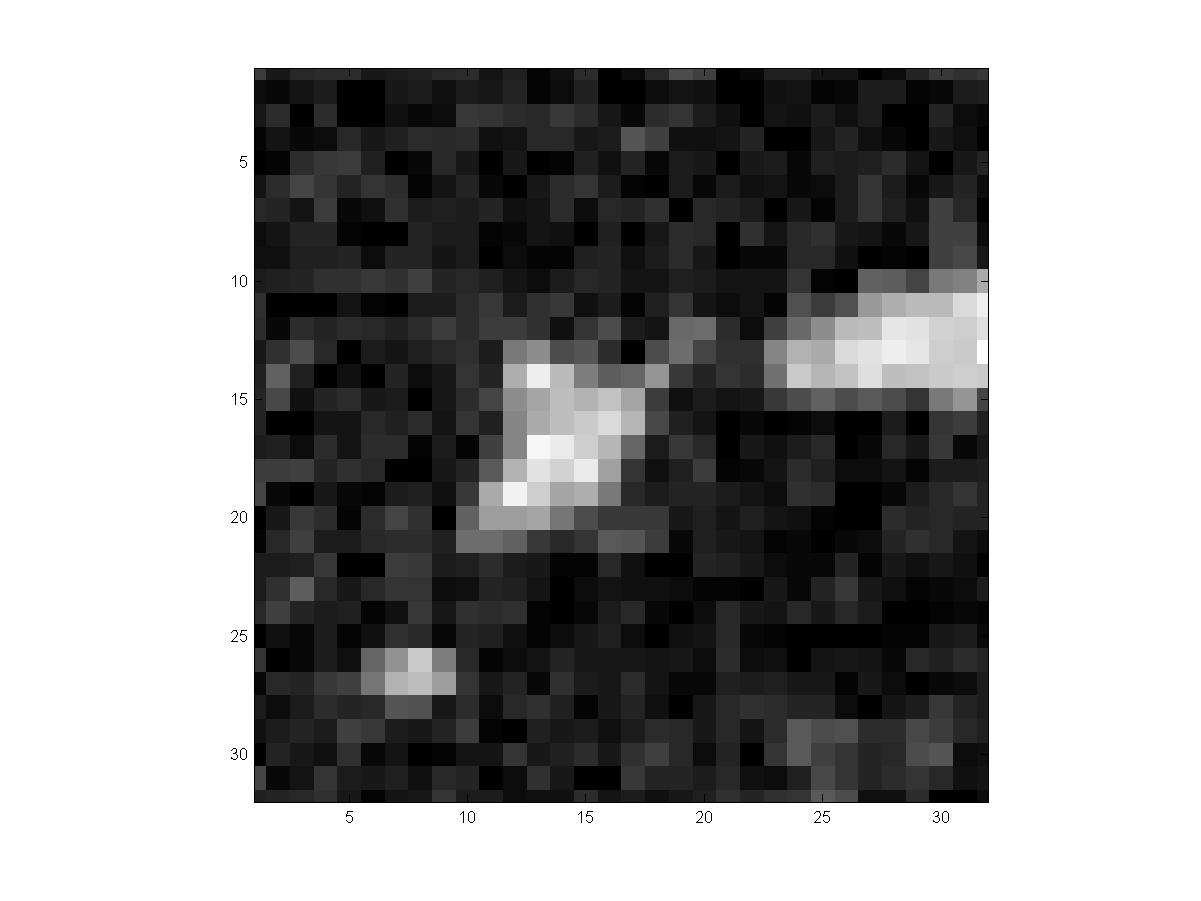
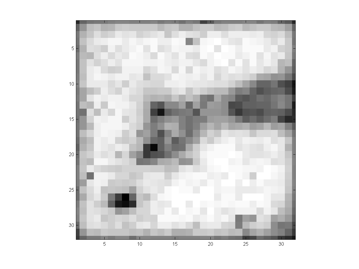
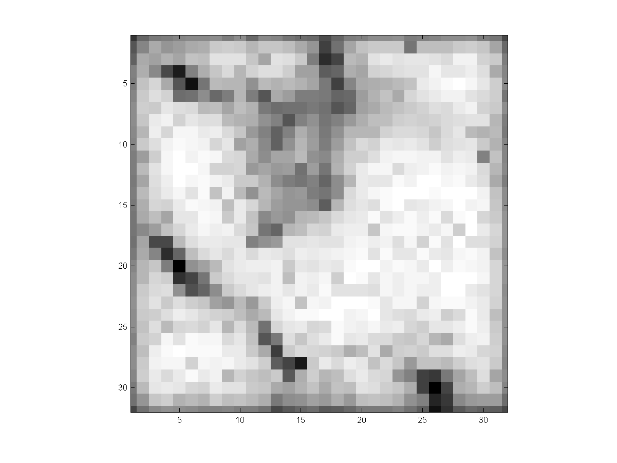
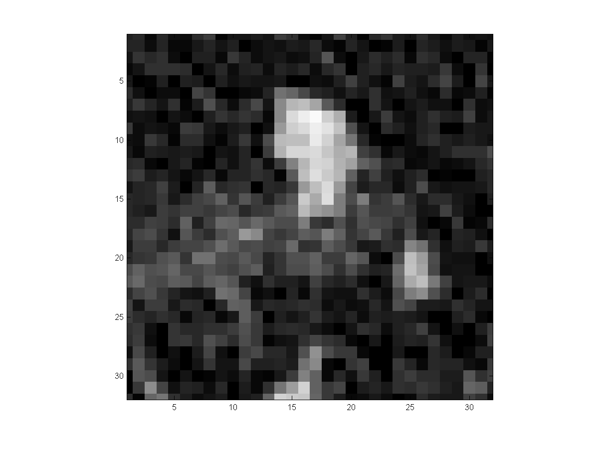
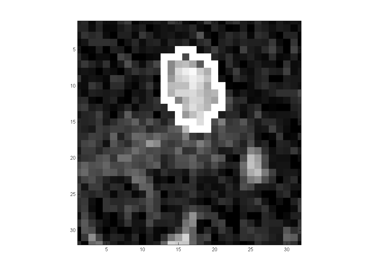
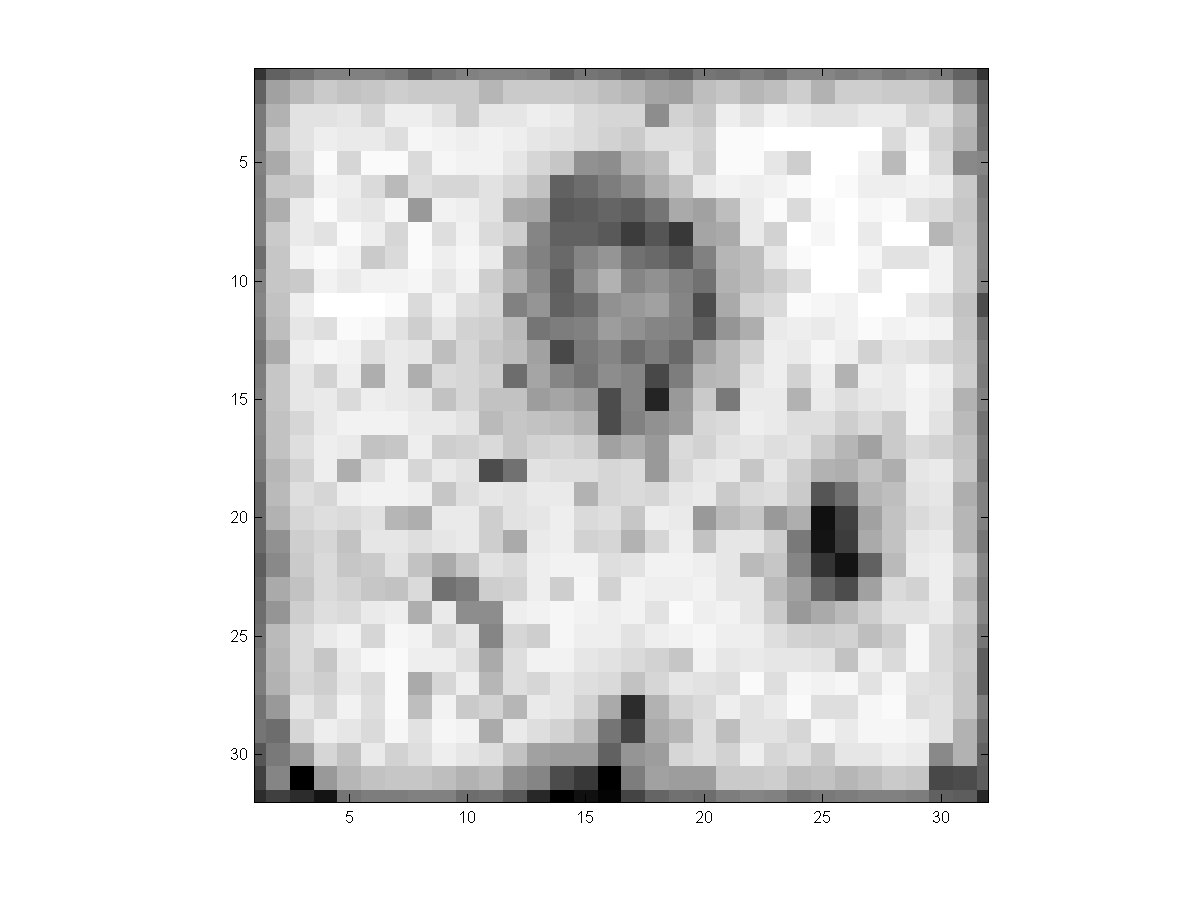
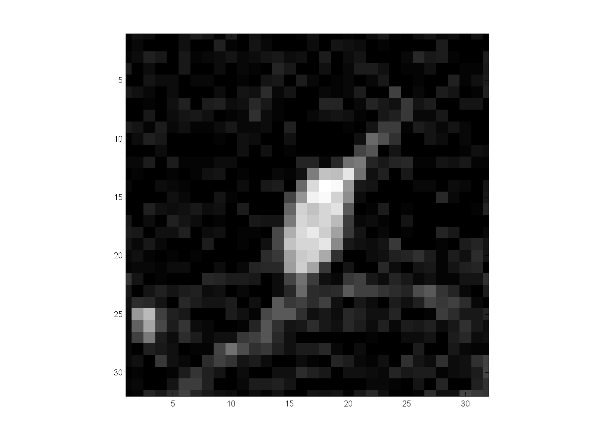
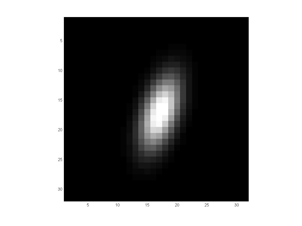
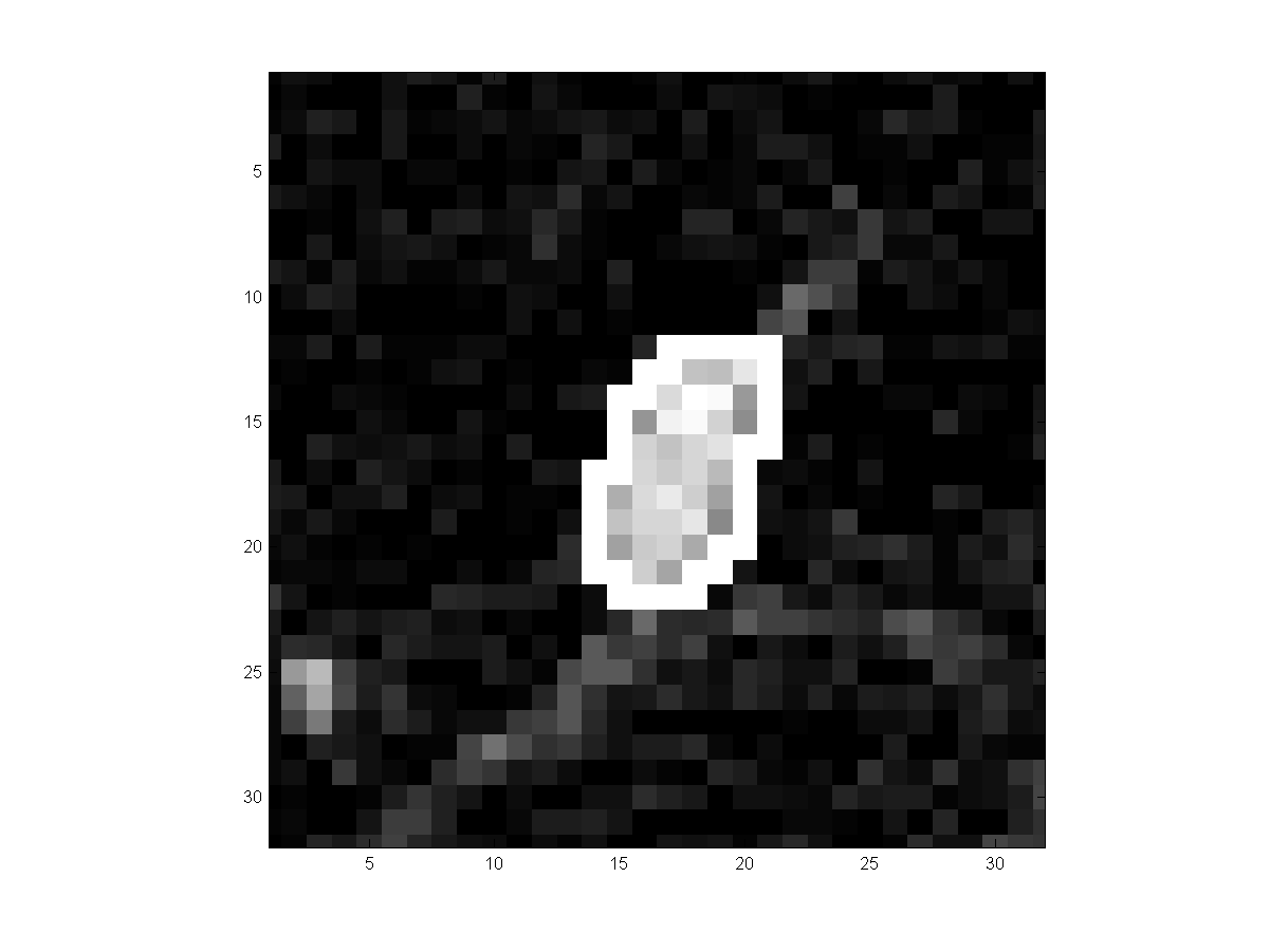
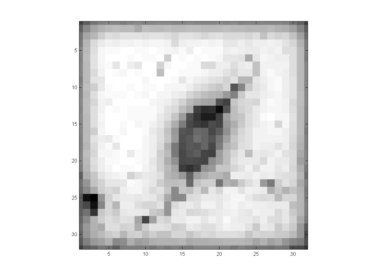
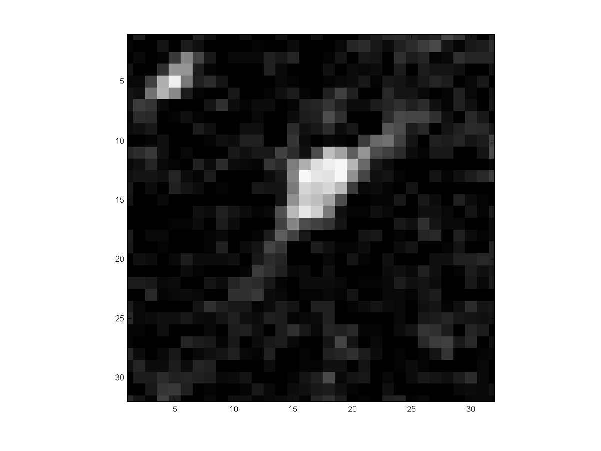
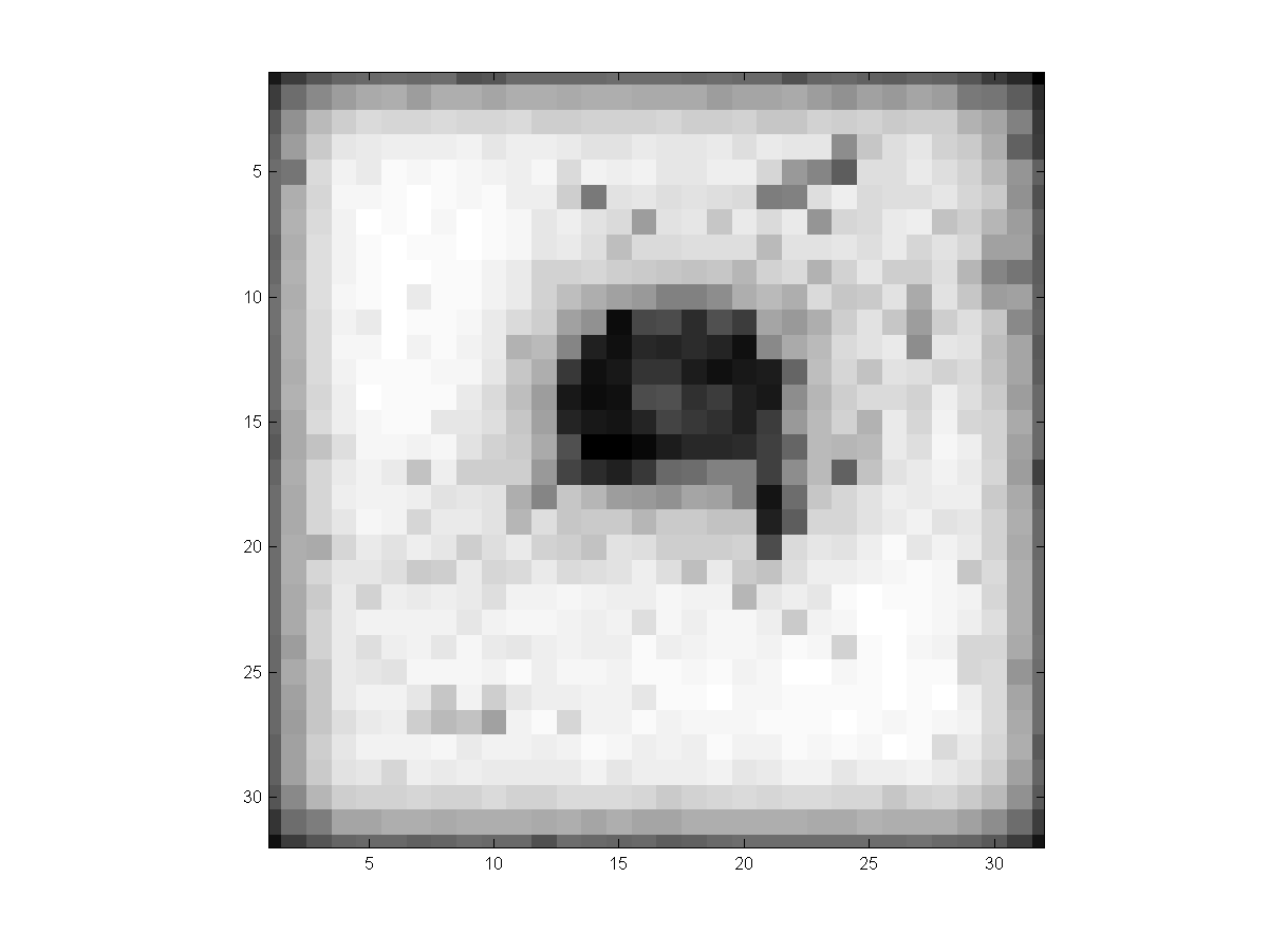
Our method's segmentation results are shown below. We use a data set of 14
patients with 77 nodules whose size ranges from 3mm to 25mm in diameter.
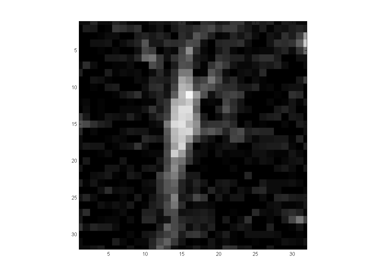
Examples are shown for i) part- or non-solid nodules, ii) pleural attached
nodules, iii) vascularized nodules, and iv) small nodules (~3mm).
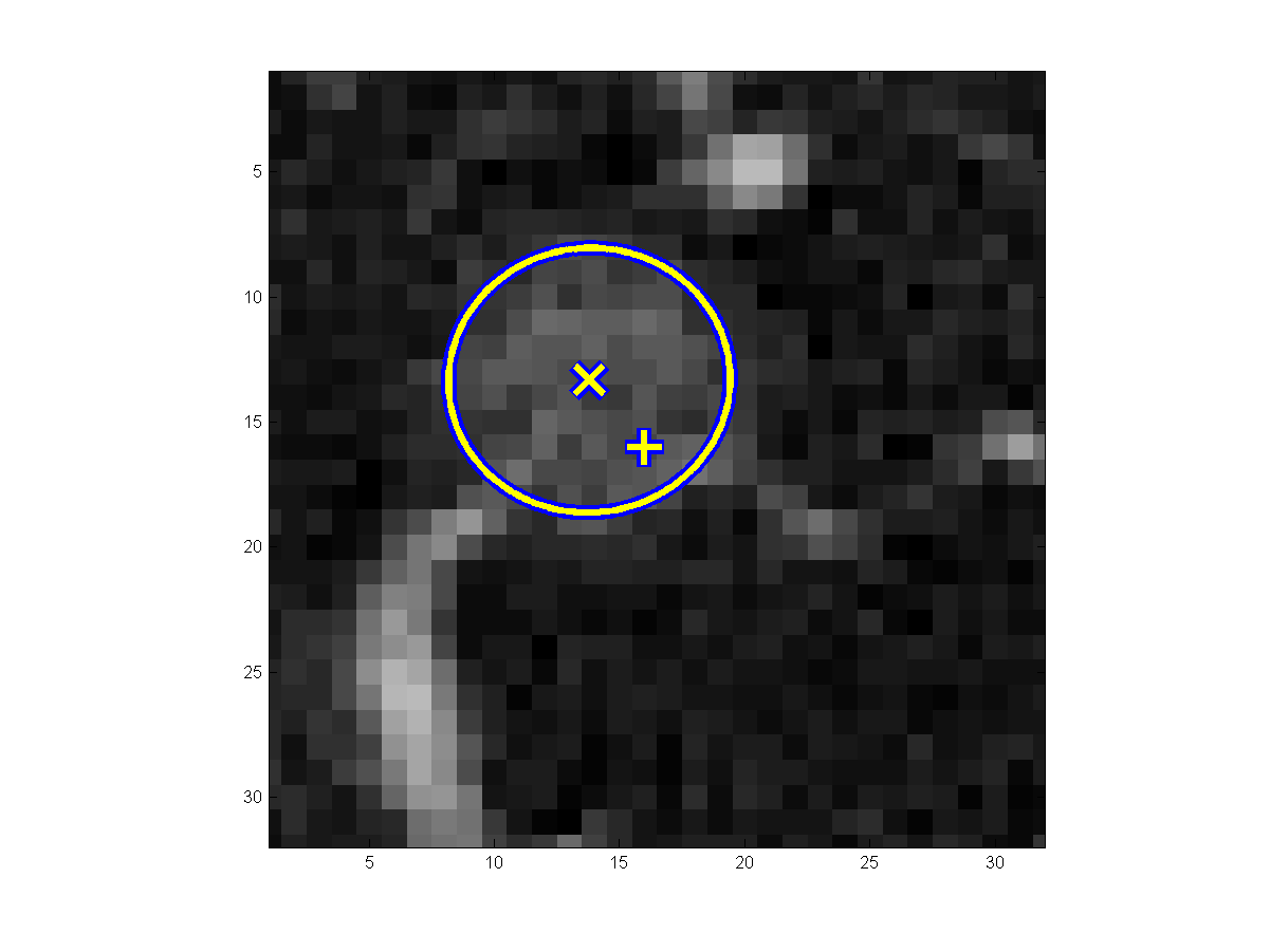
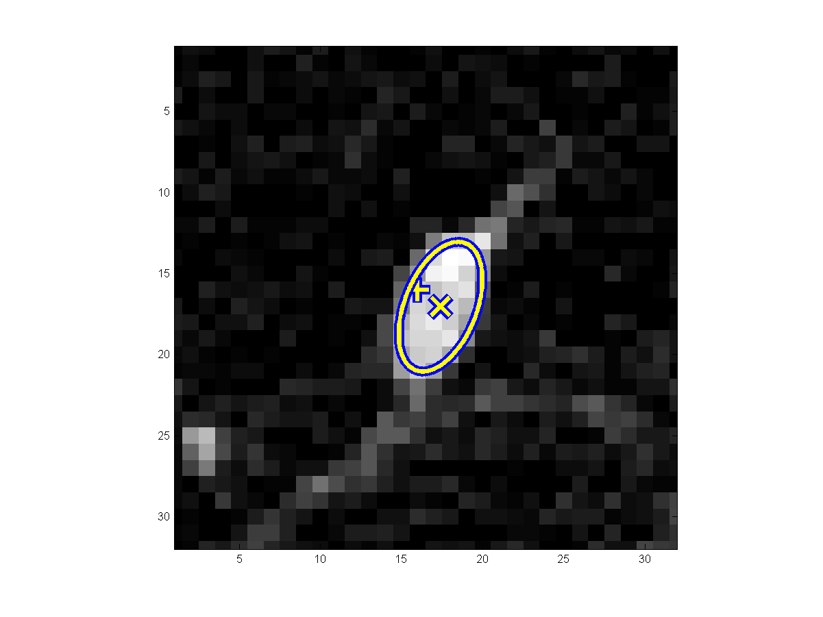
For
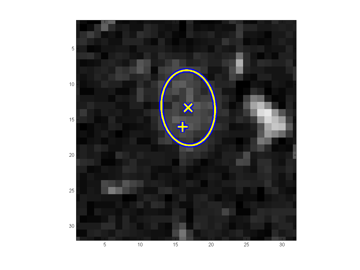
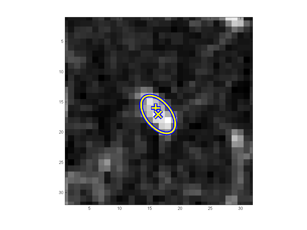
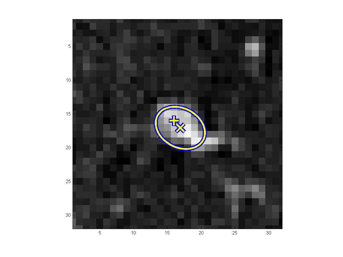
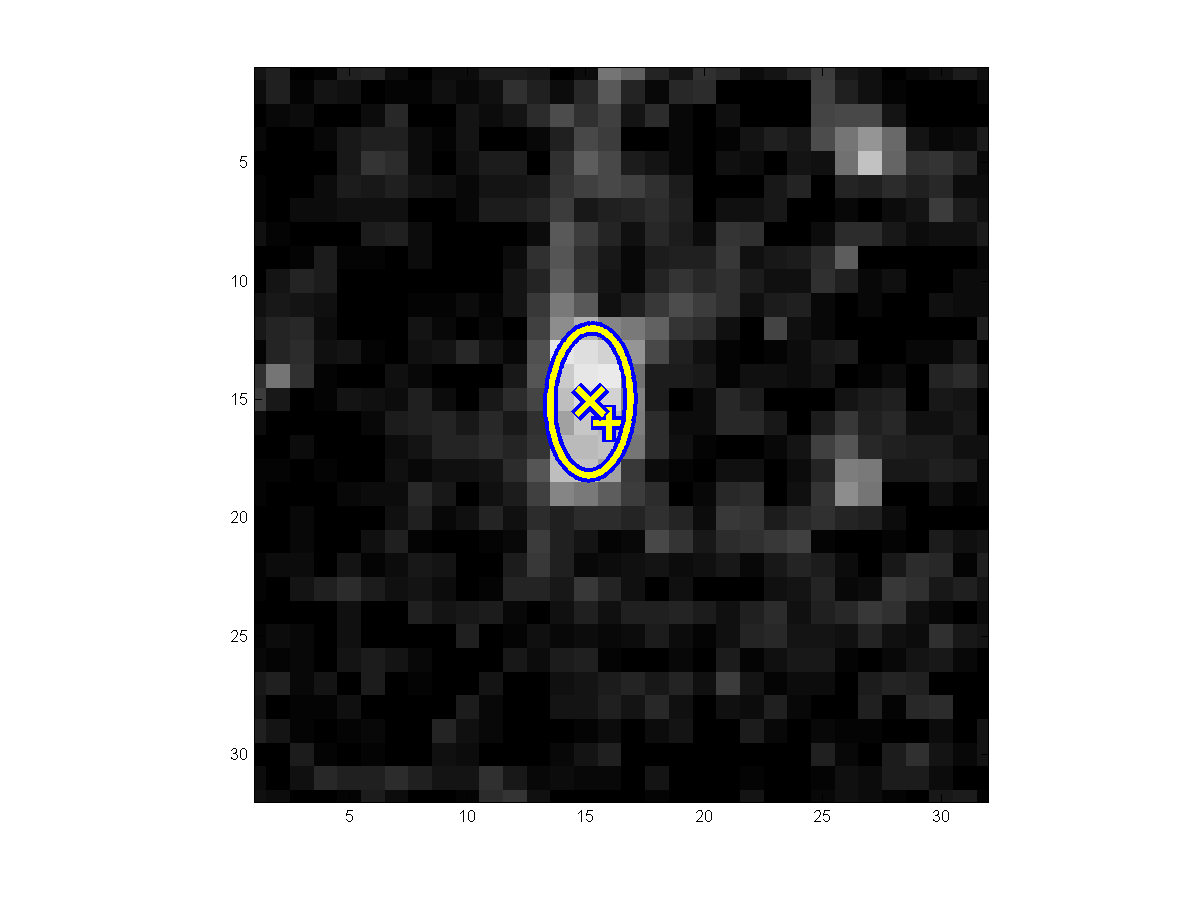
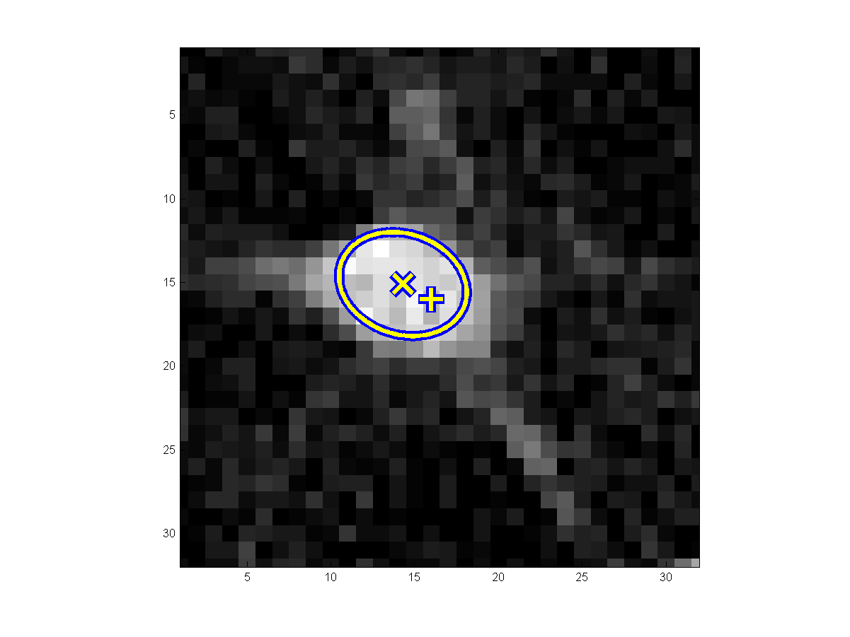
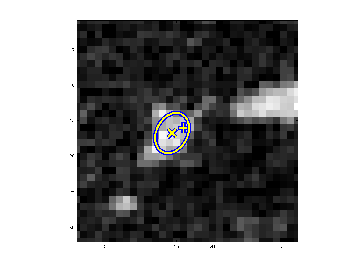
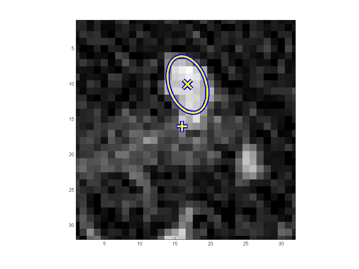
comparison, the results by a) the parametric (anisotropic Gaussian/ellipsoidal)
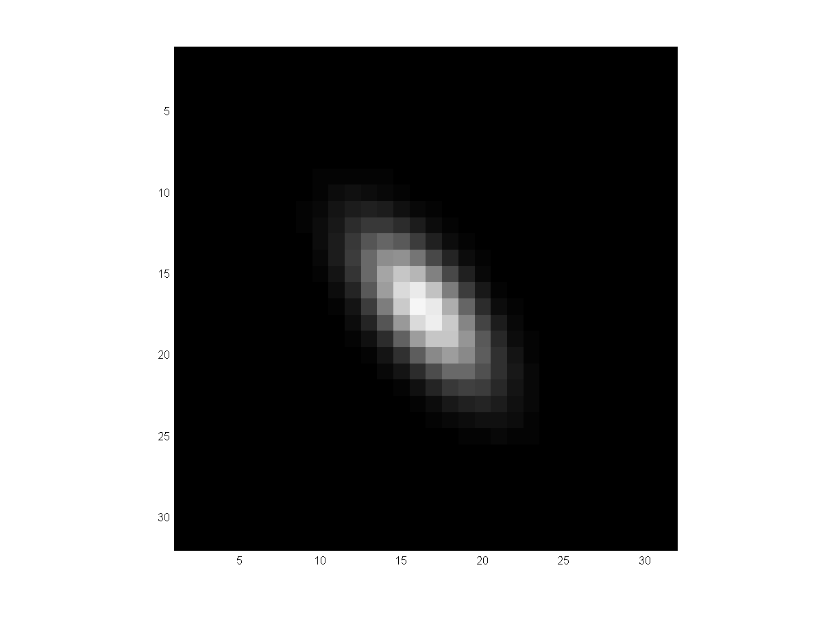
model fitting (2nd columns) and b) the non-parametric 4D joint space
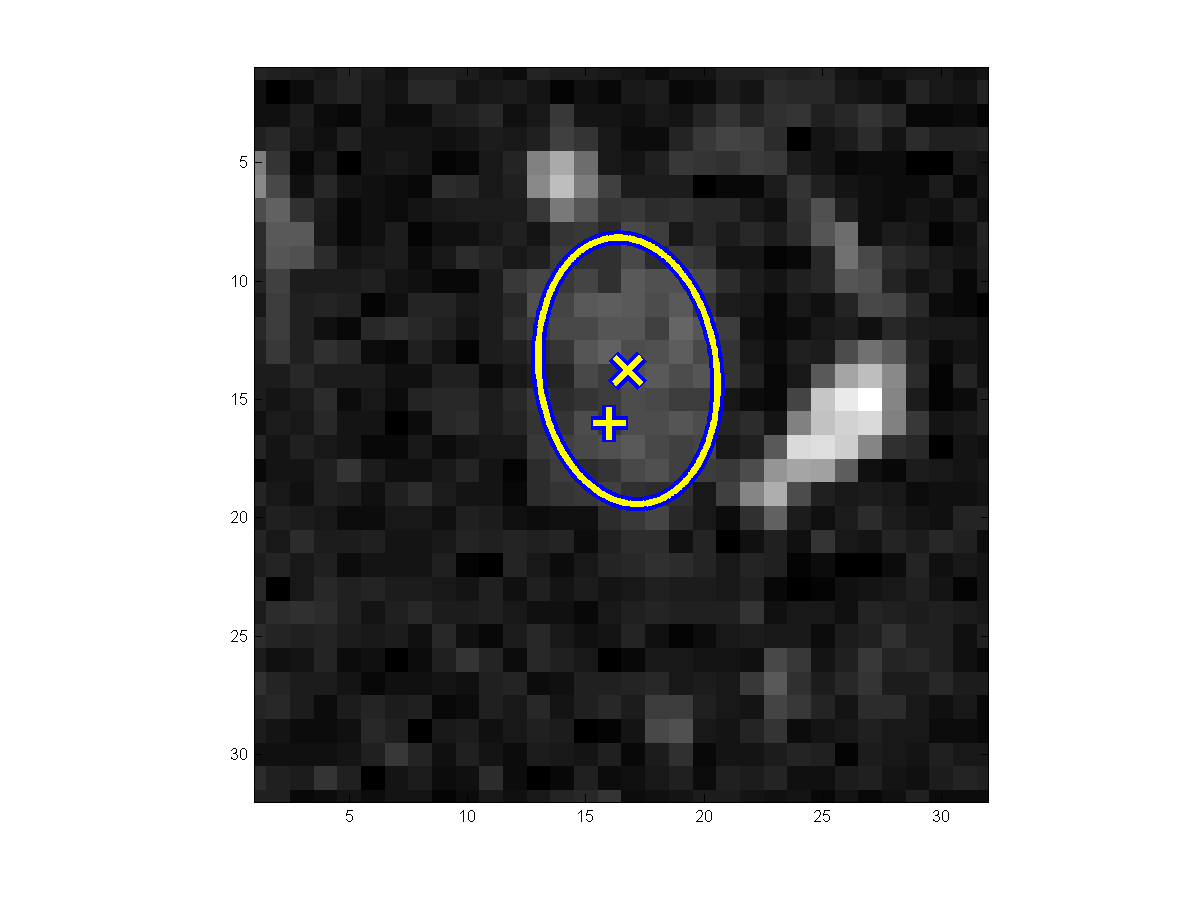
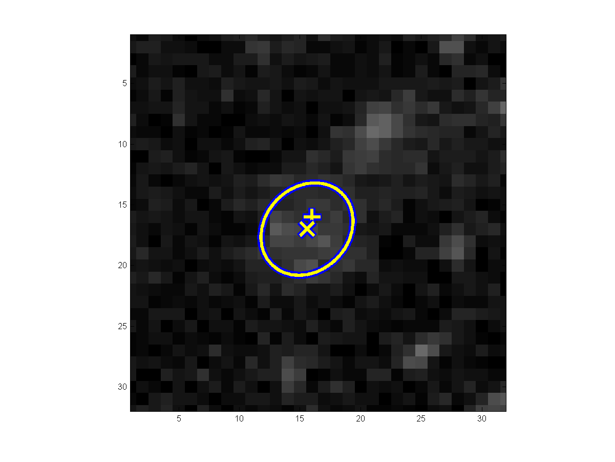
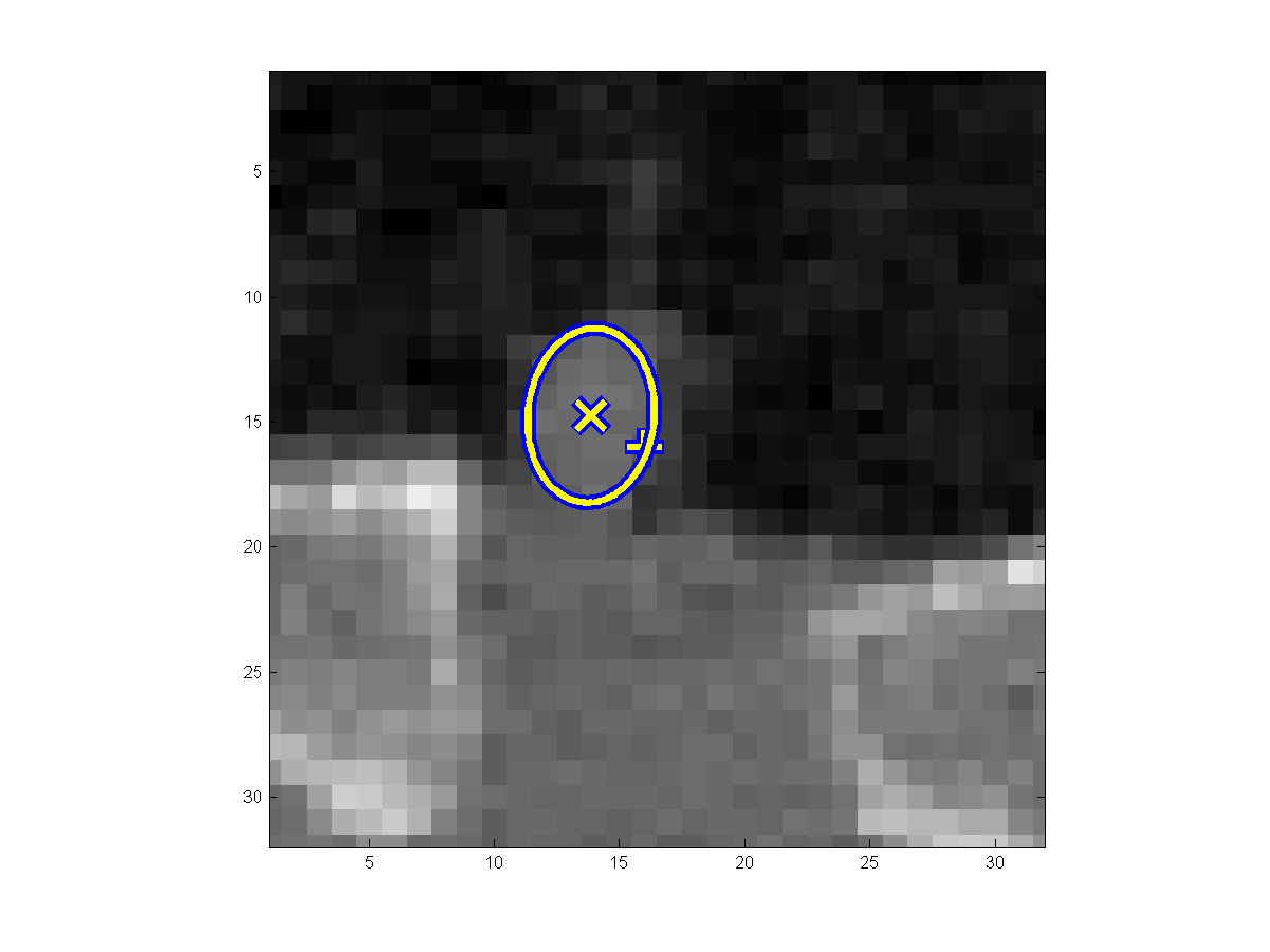
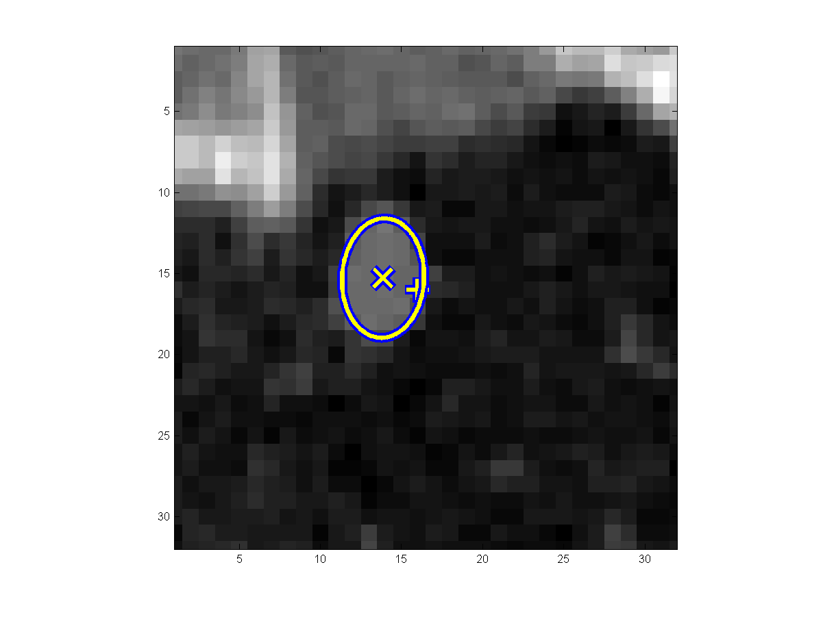
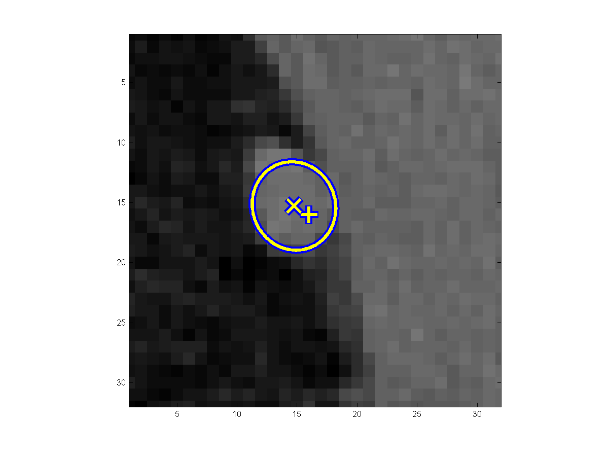
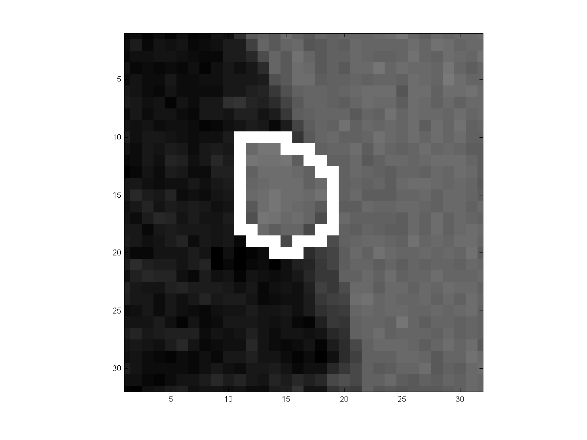
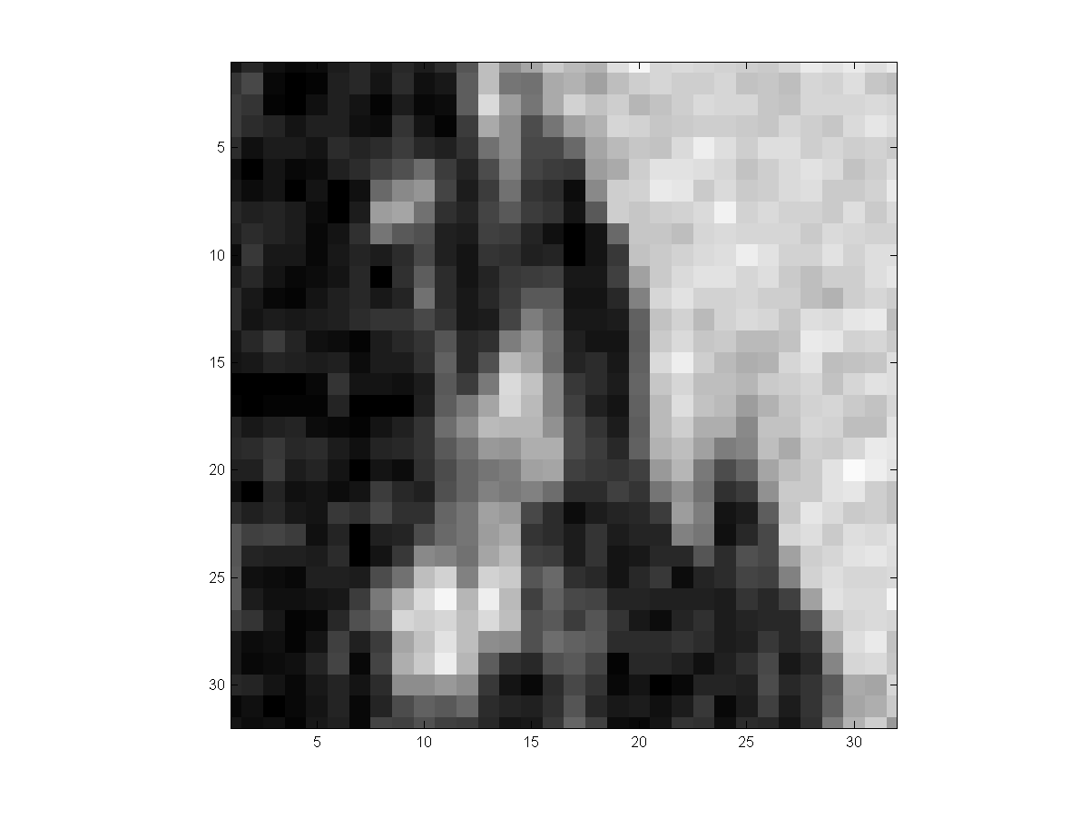
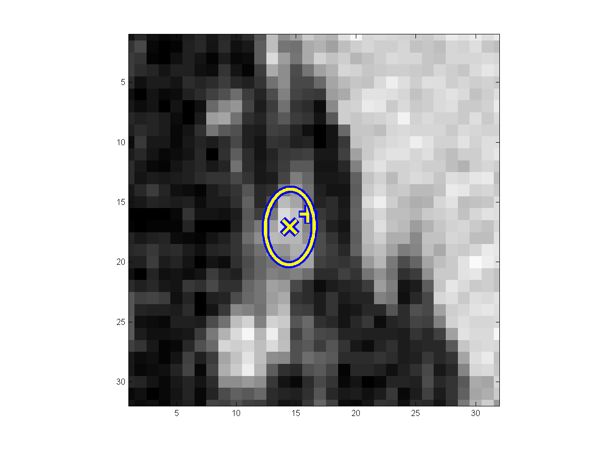
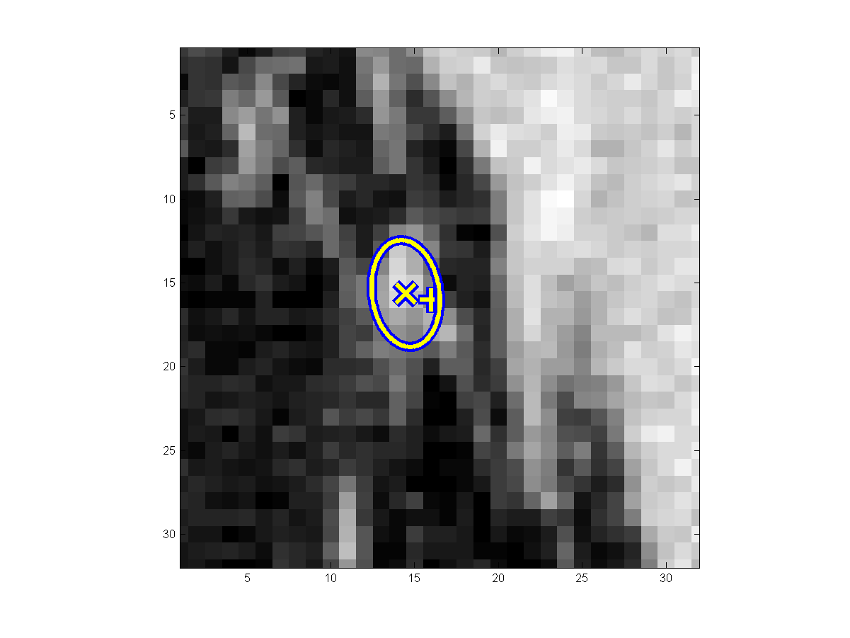
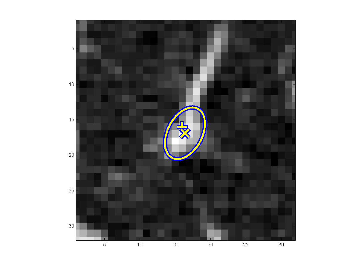
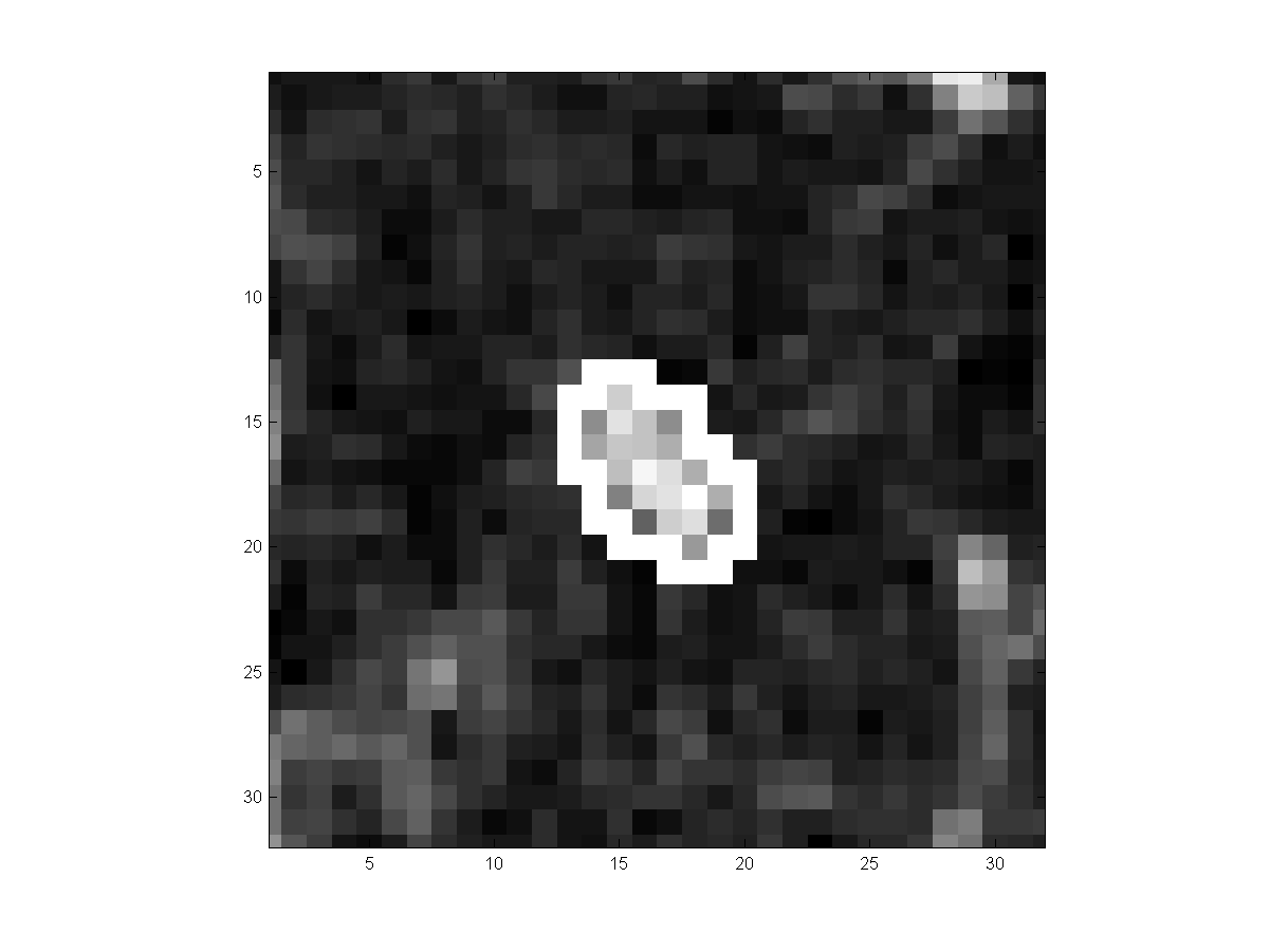
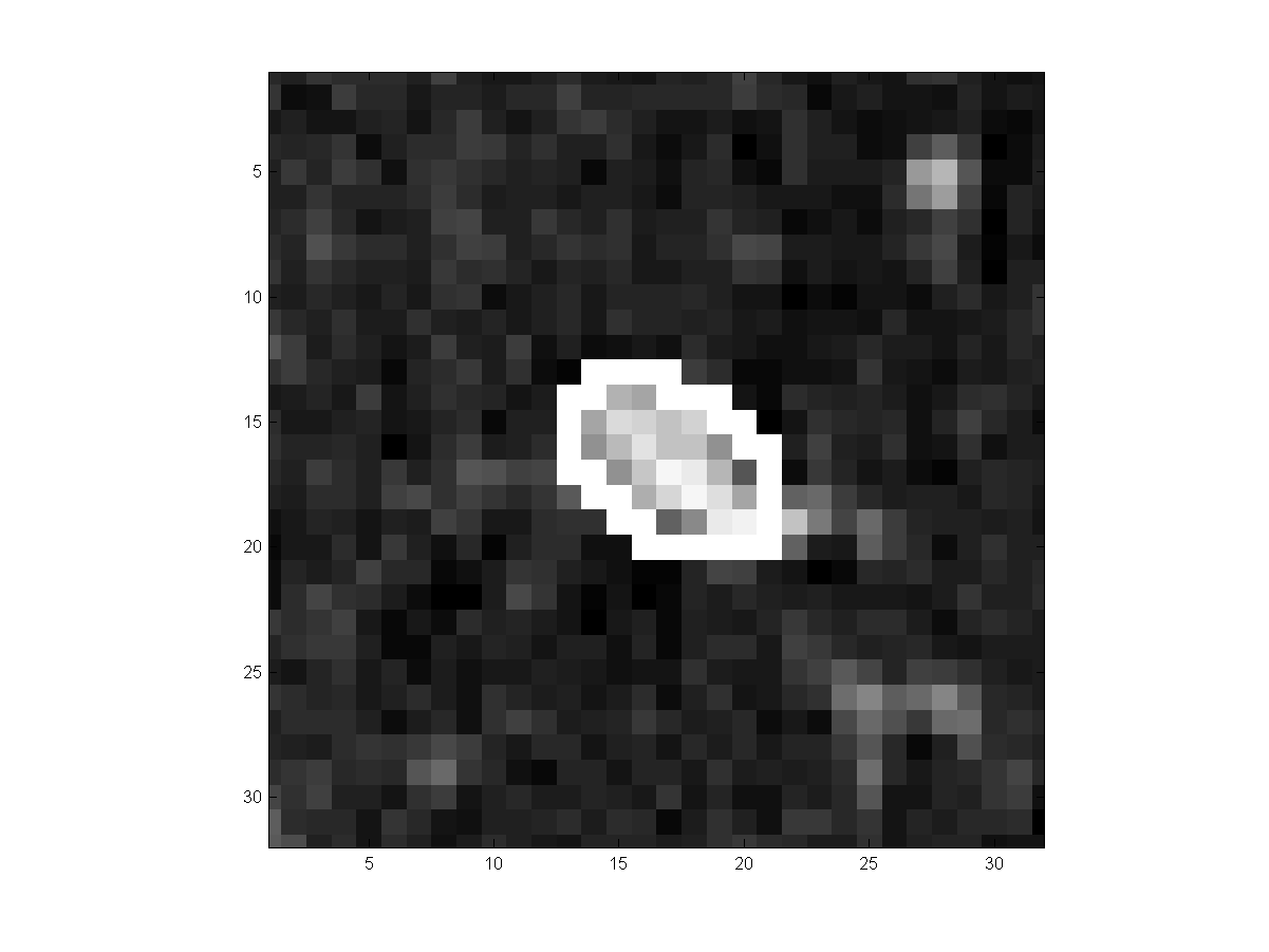
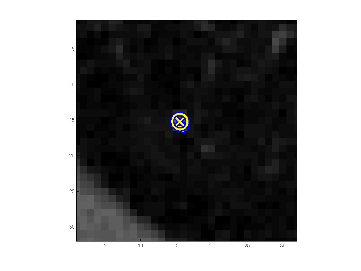
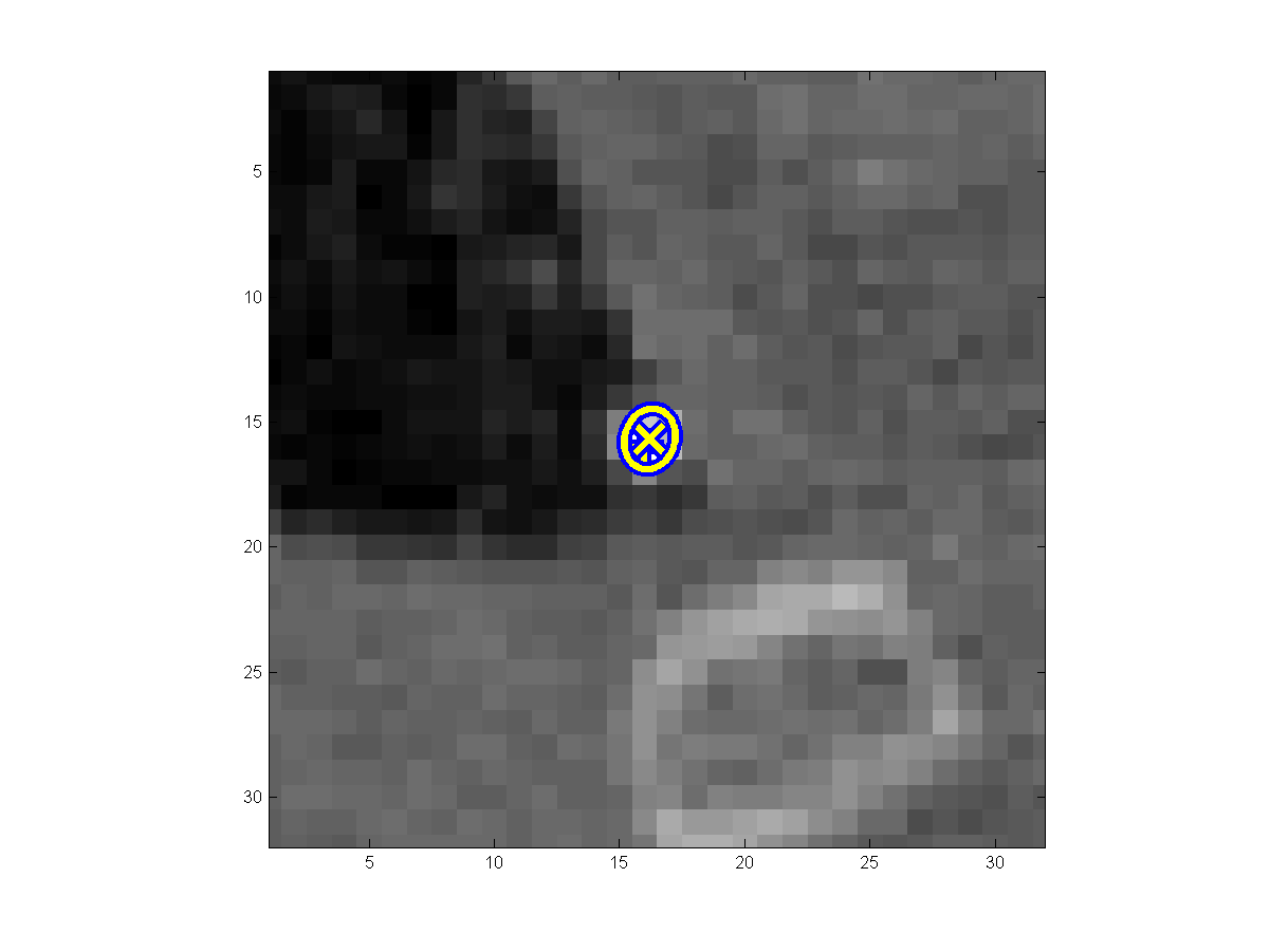
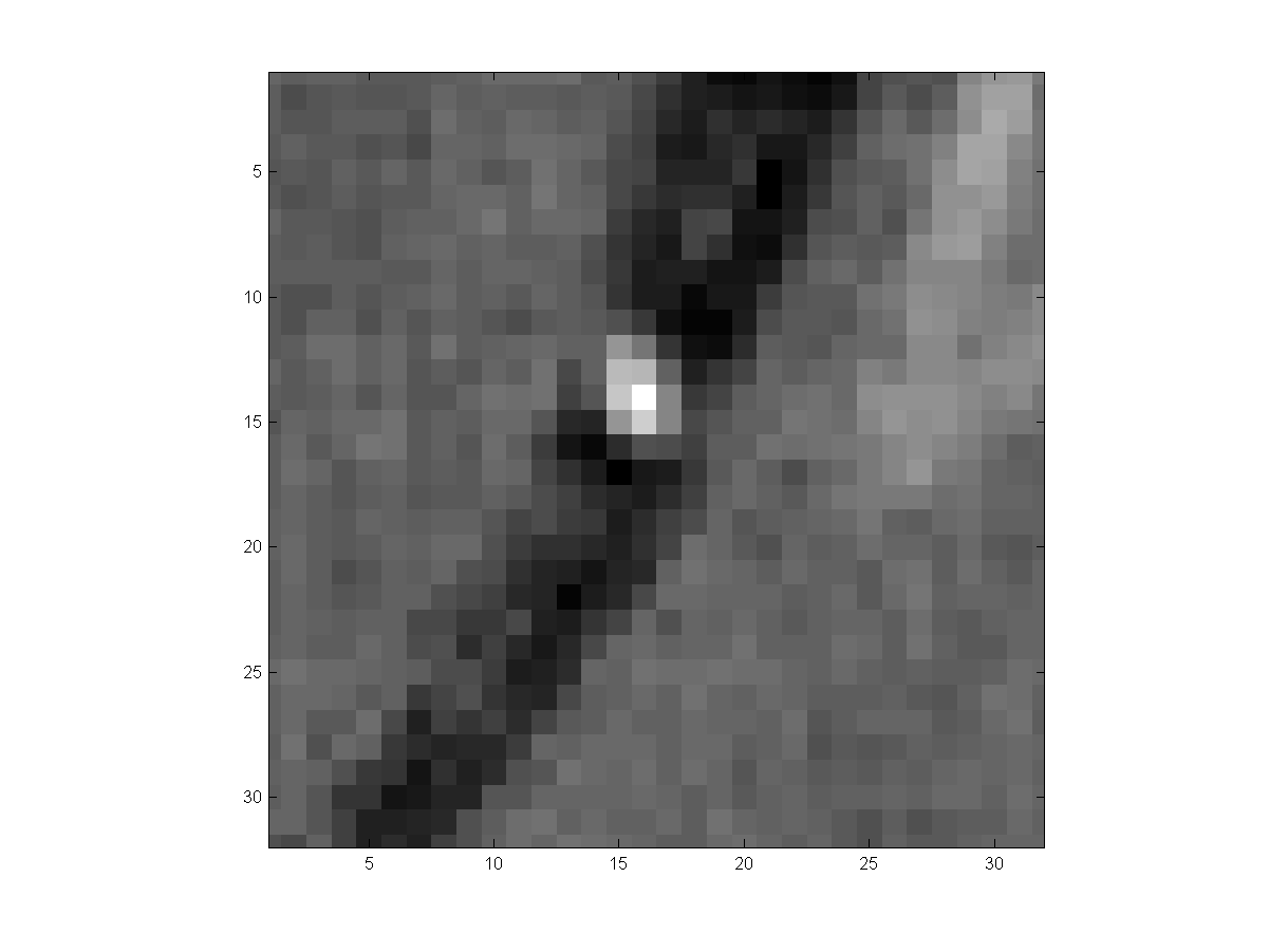
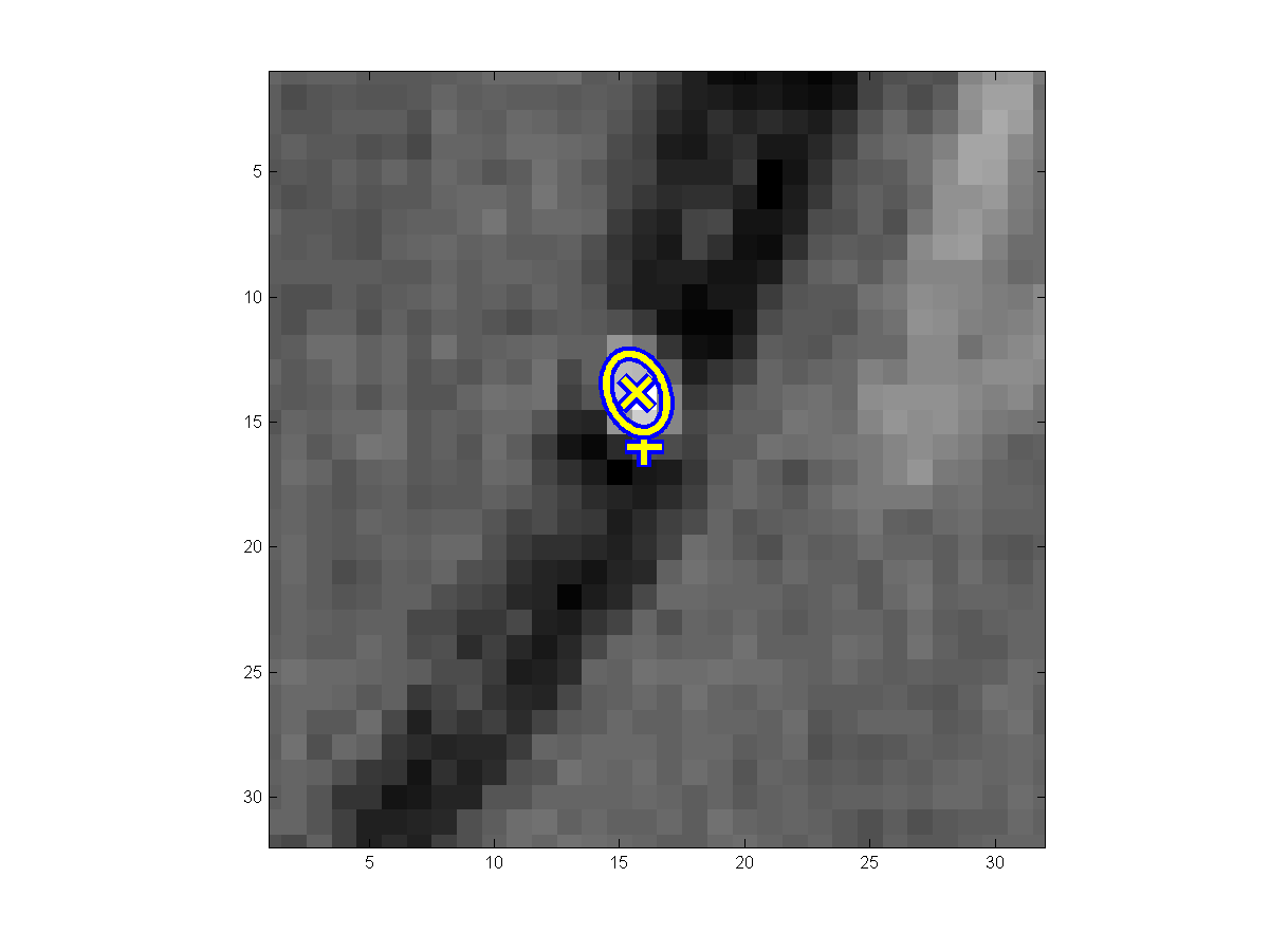
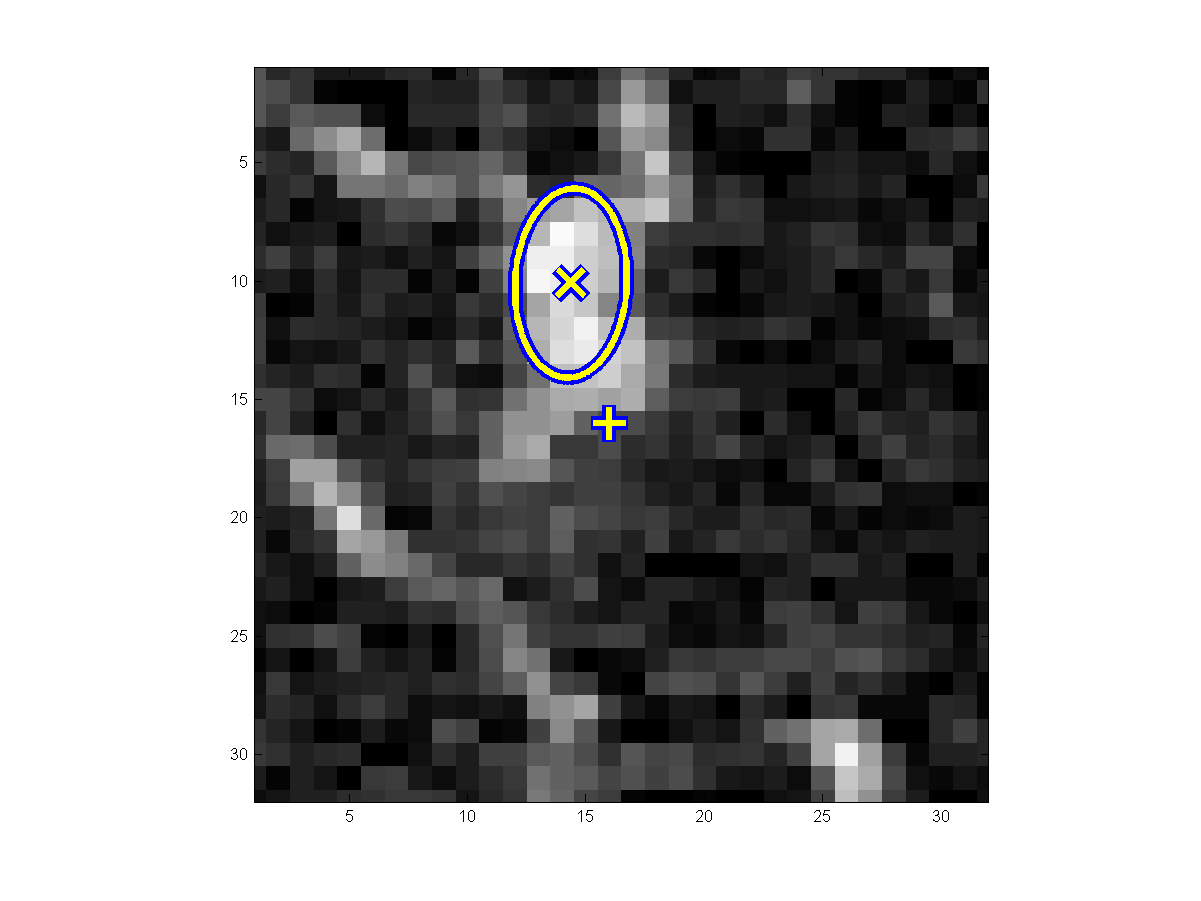
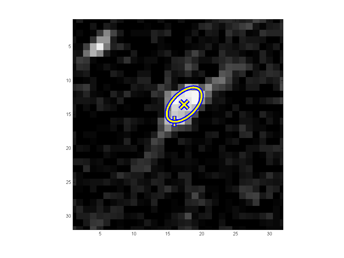
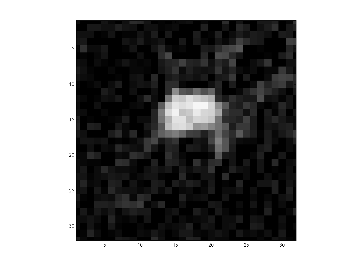
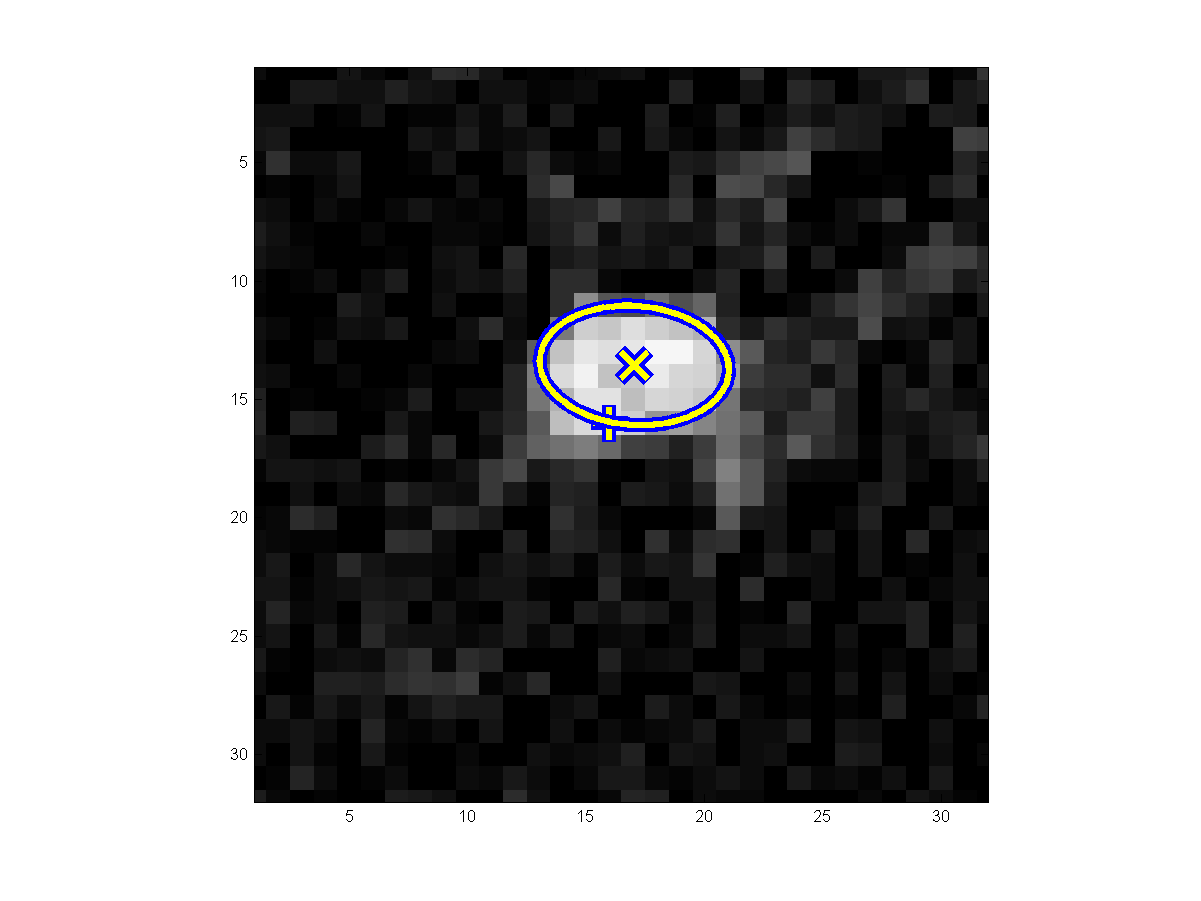
segmentation (3rd columns) are shown for each nodule cases. In the 2nd columns,
"+" and "x" indicate the marker location and tumor center estimate,
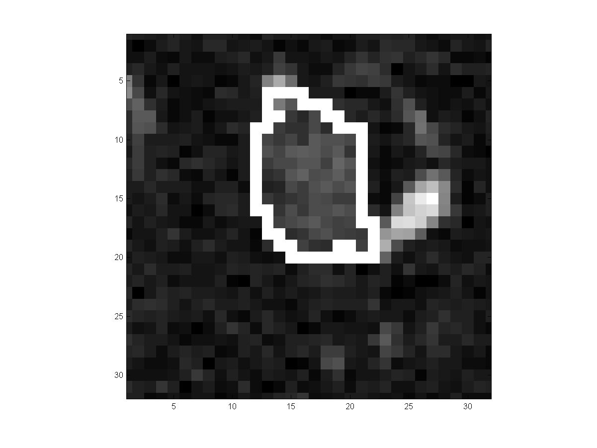
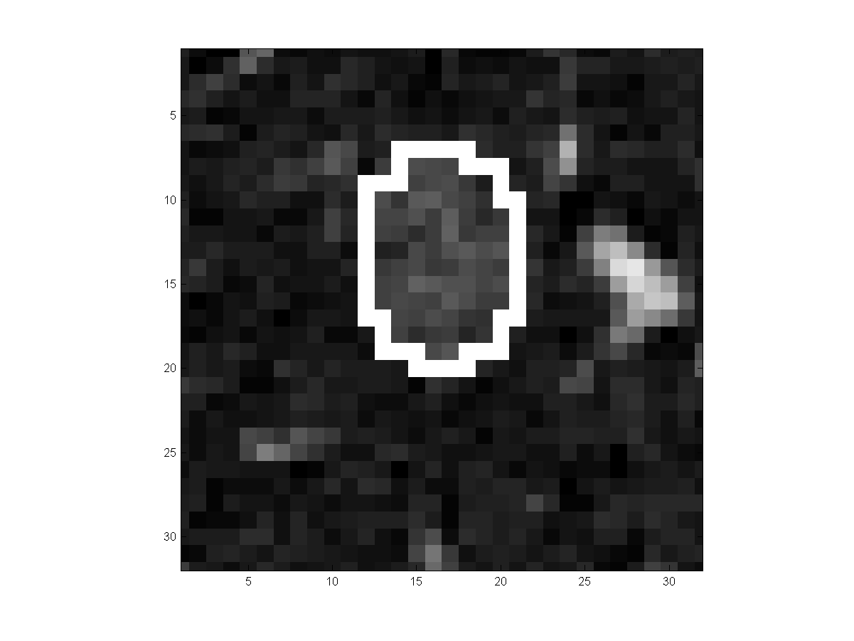
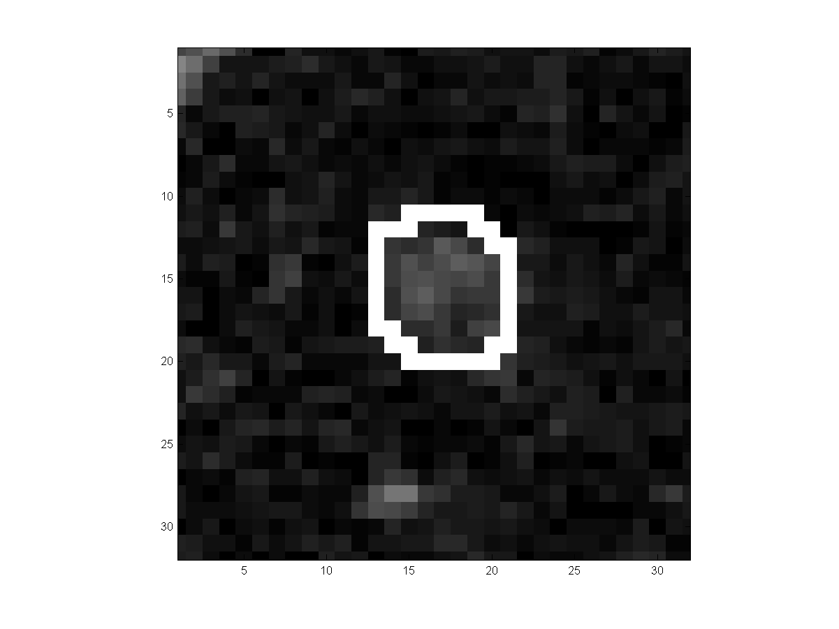
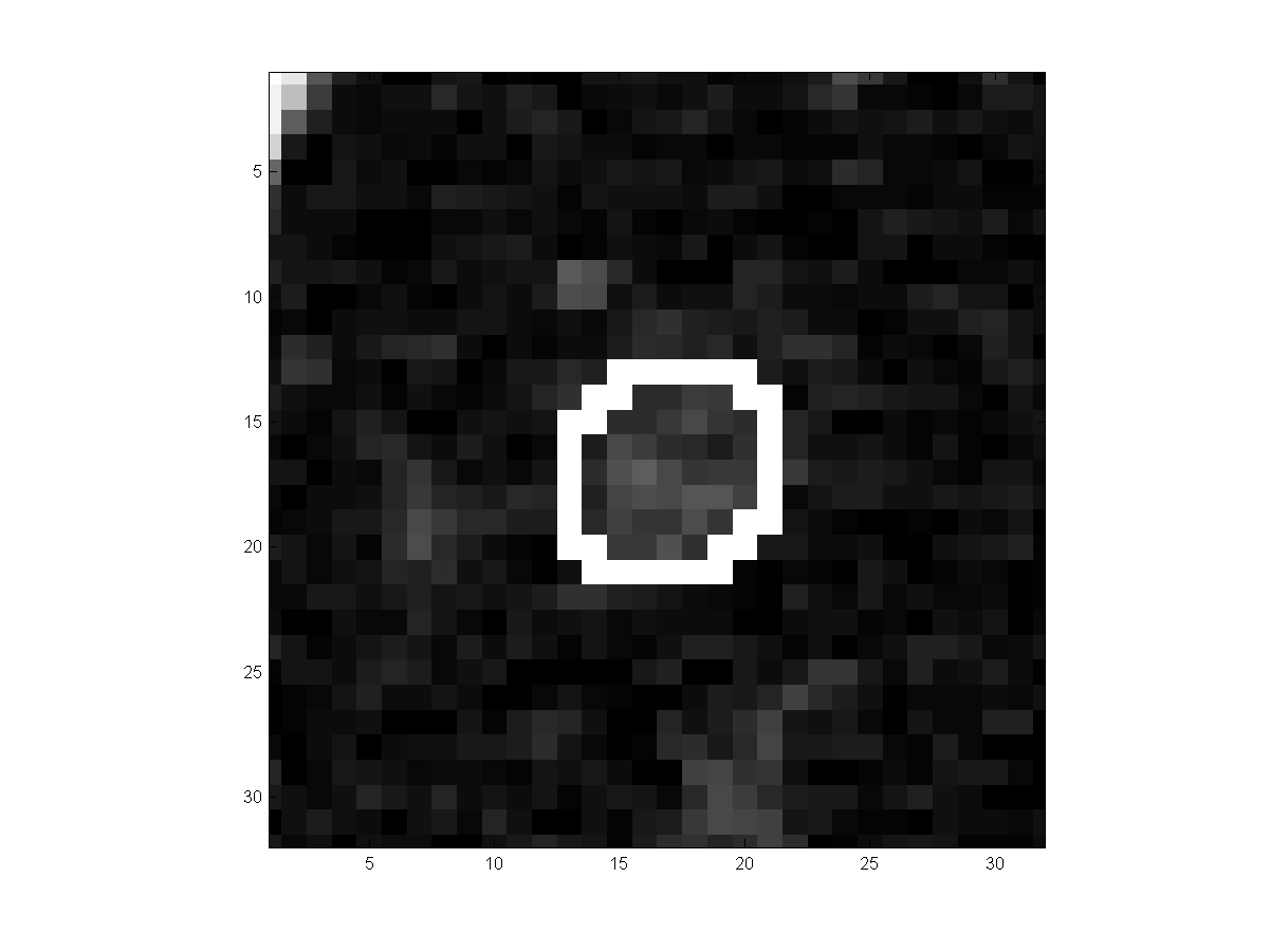
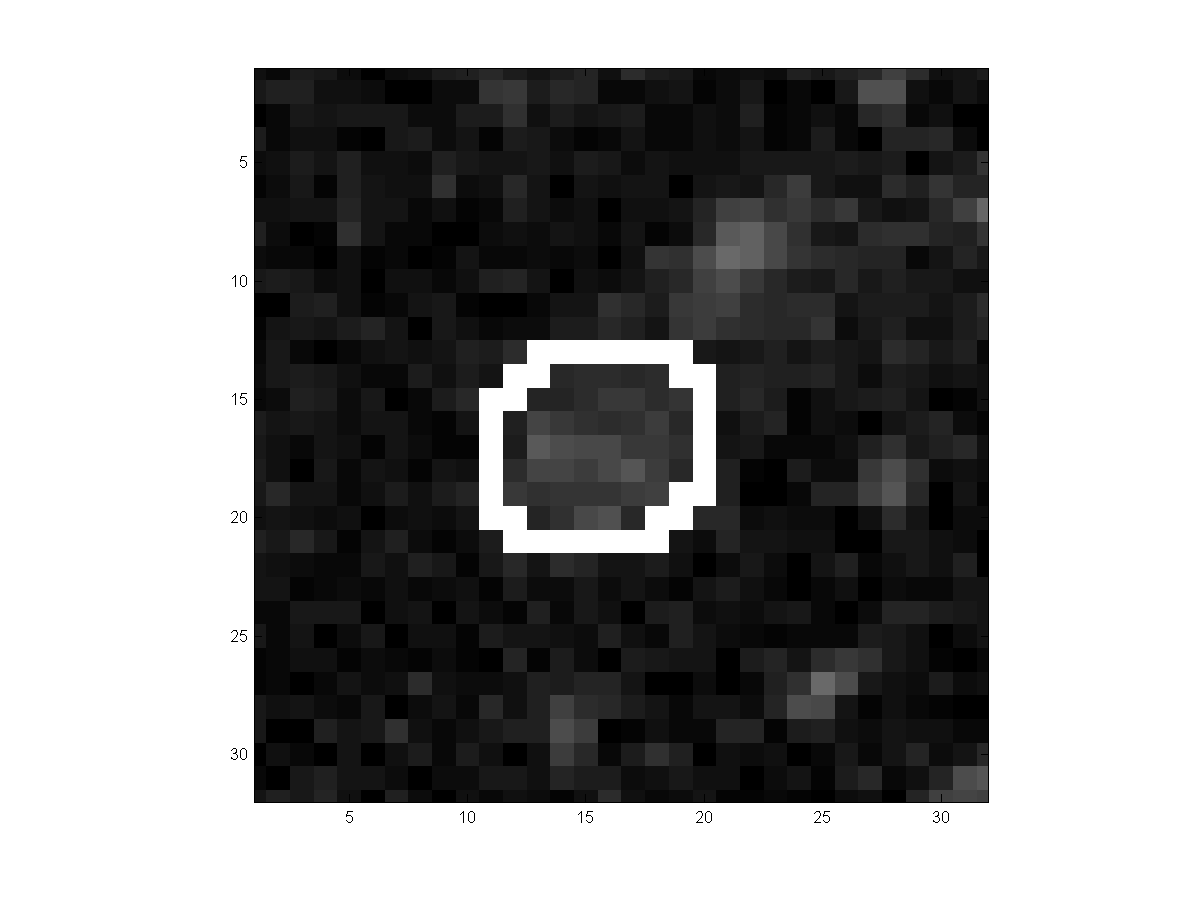
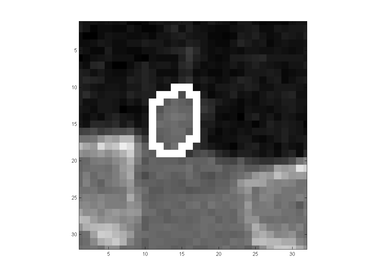
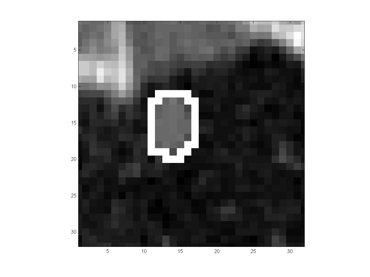
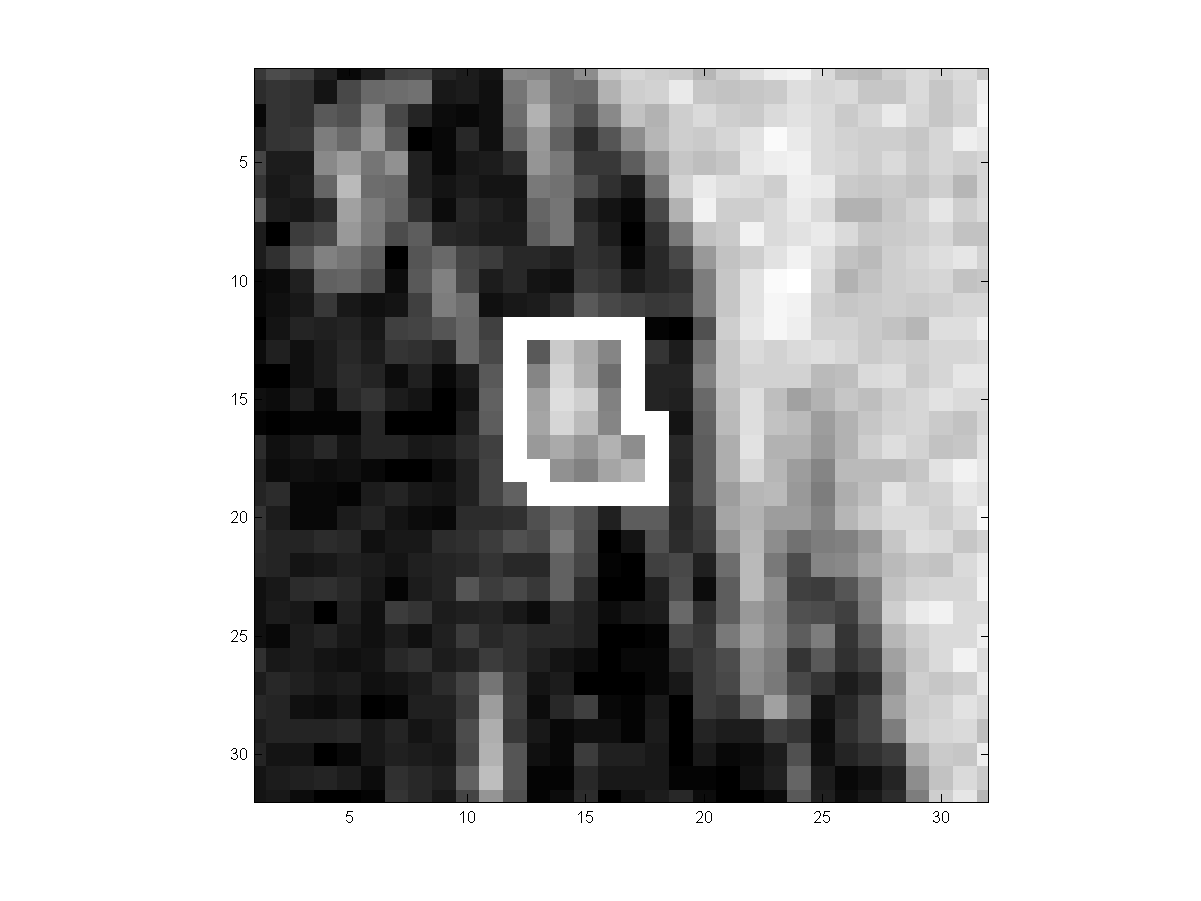
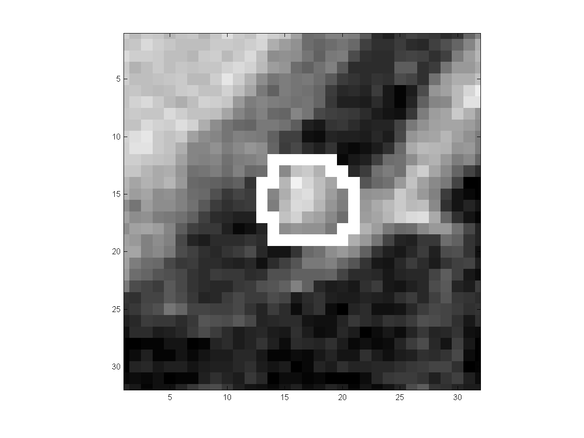
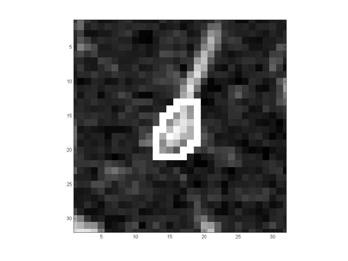
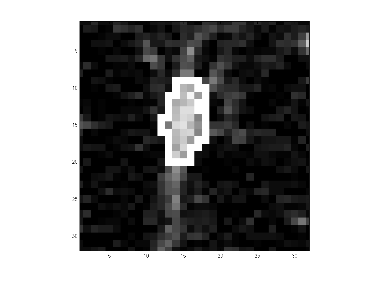
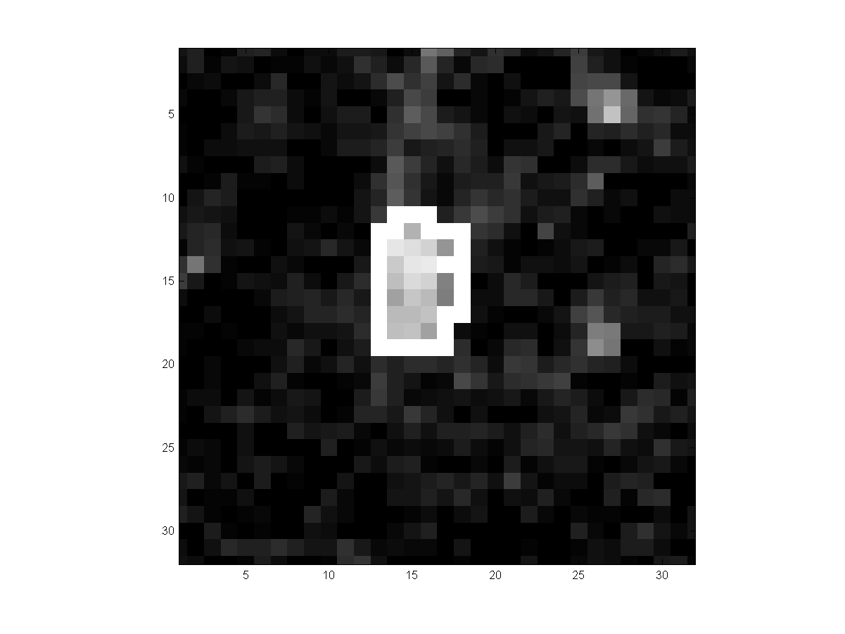
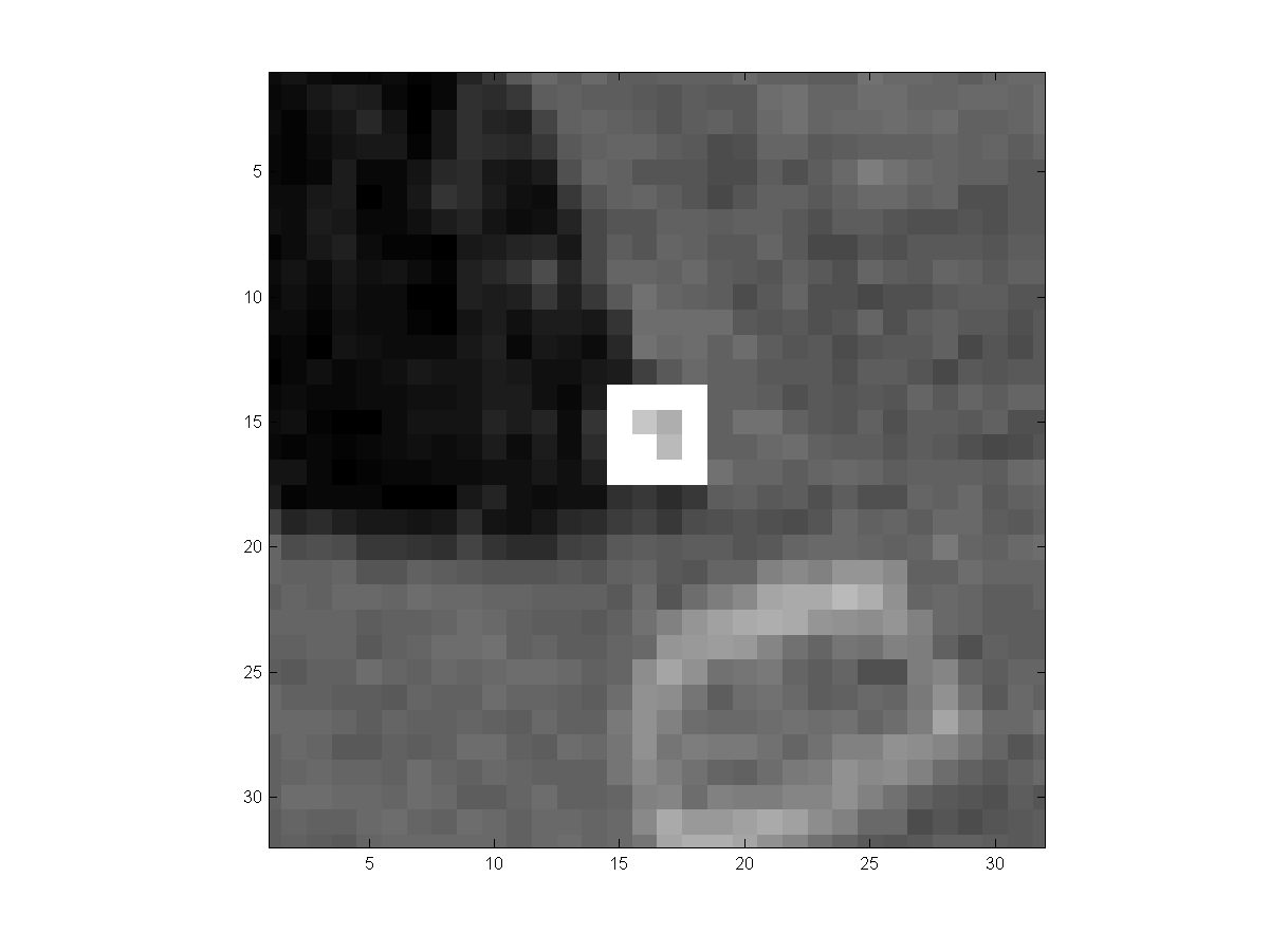
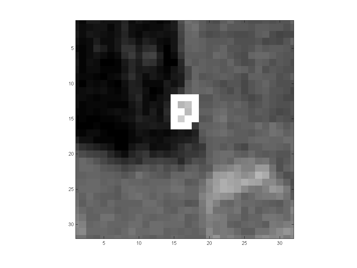
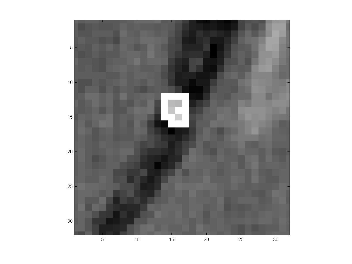
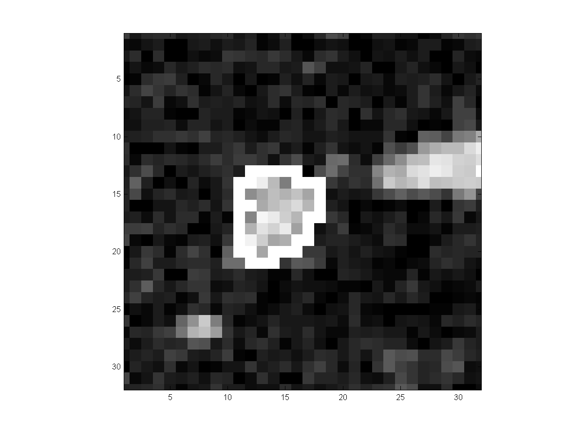
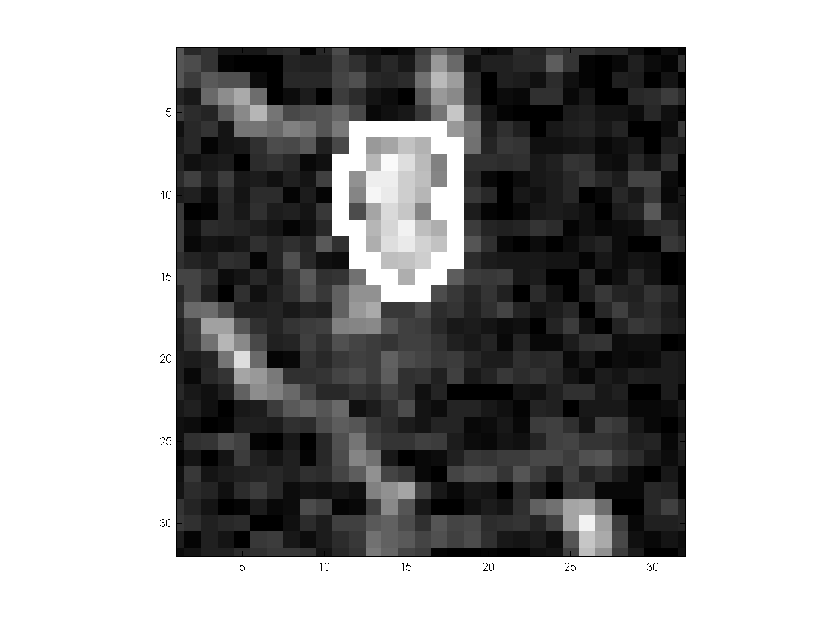
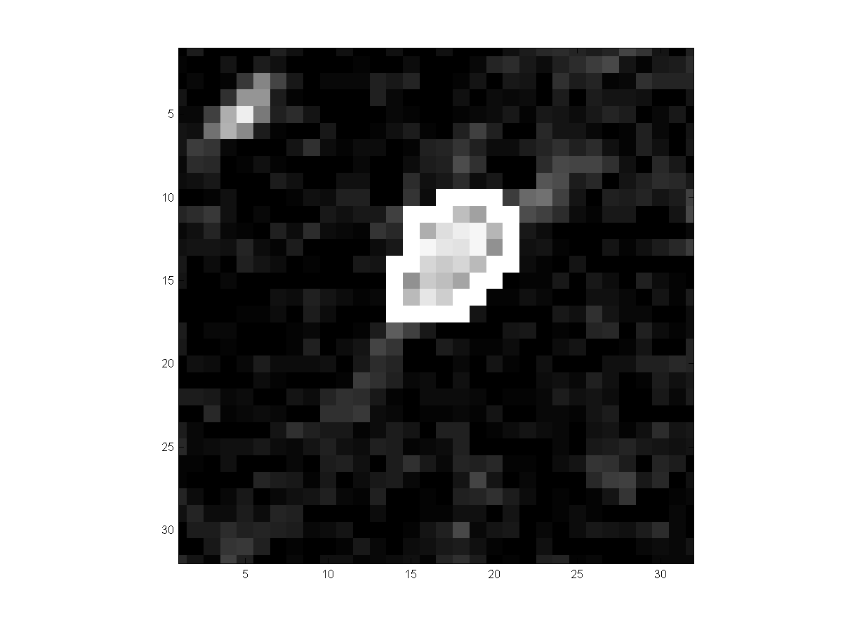
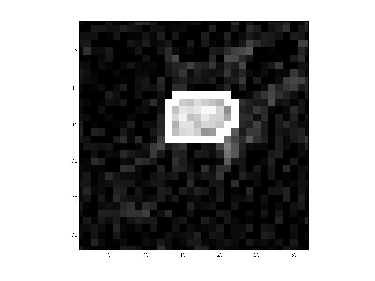
respectively. In the 4th columns, the white pixels outline the outside boundary
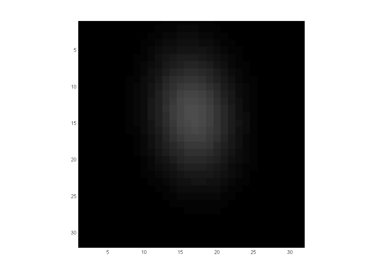
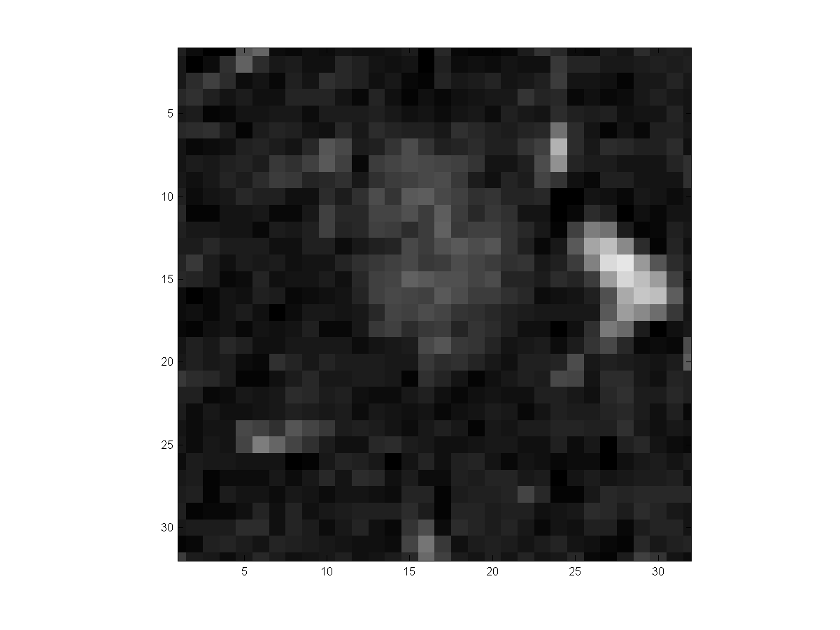
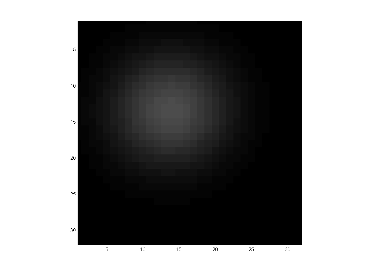
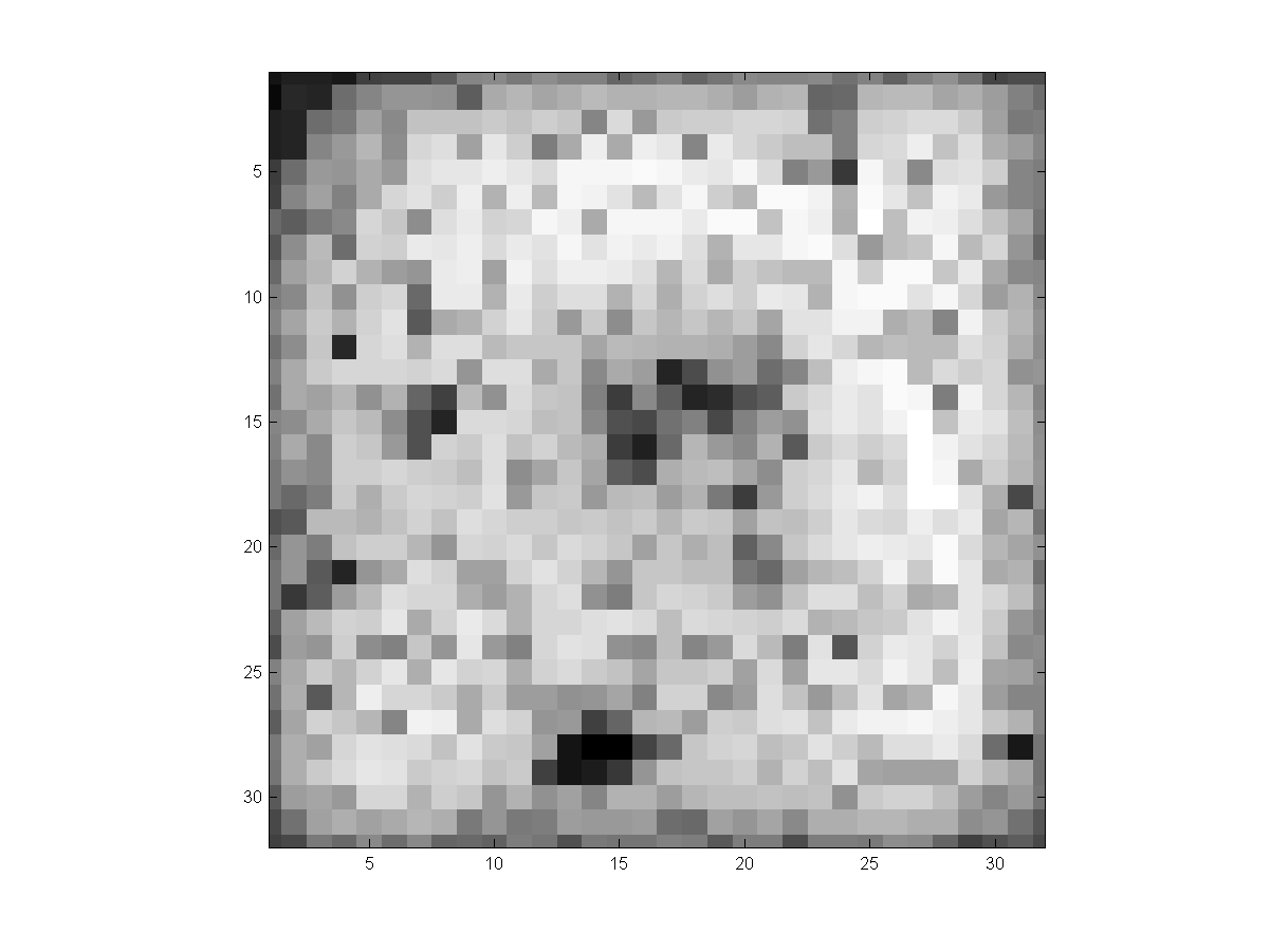
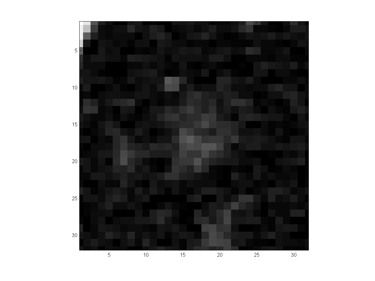
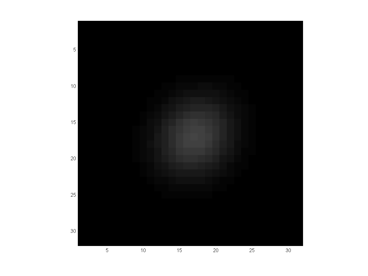
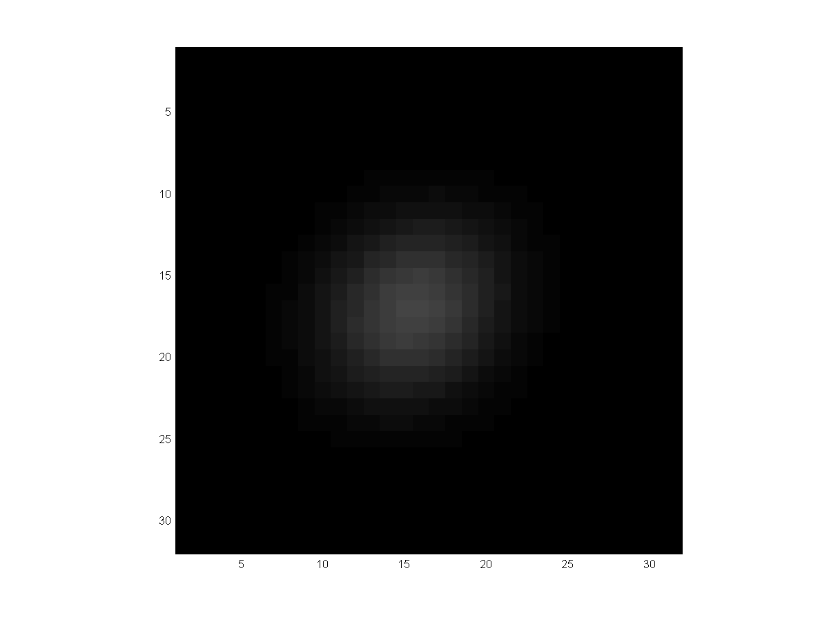
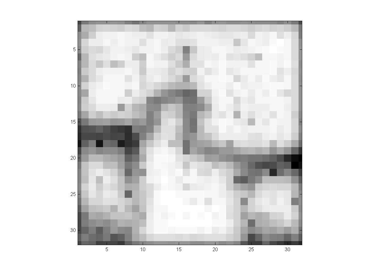
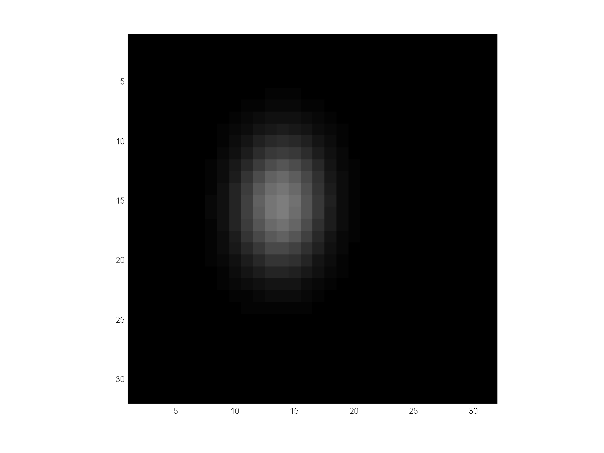
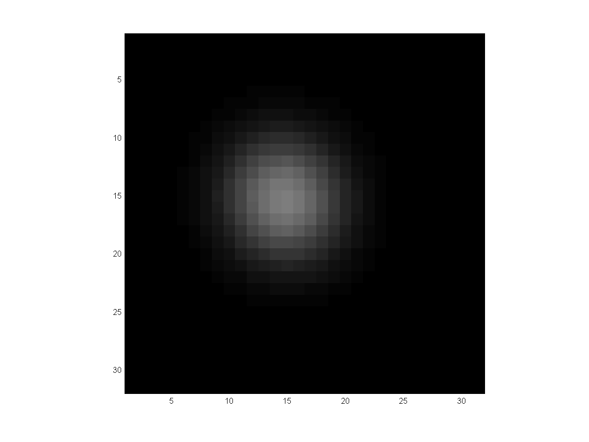
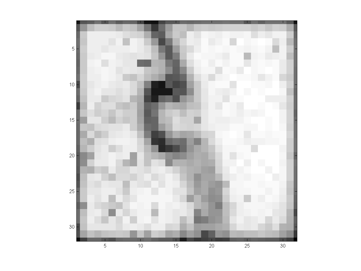
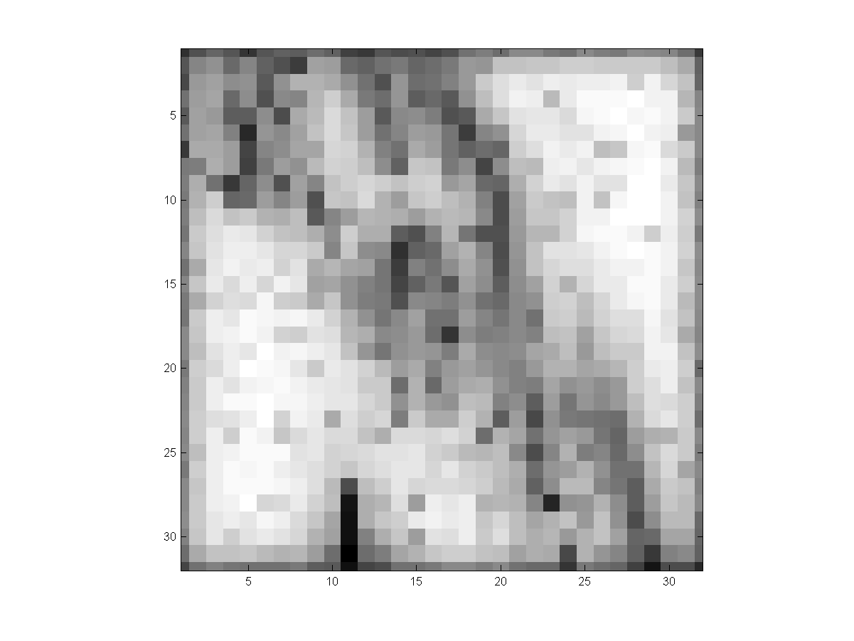
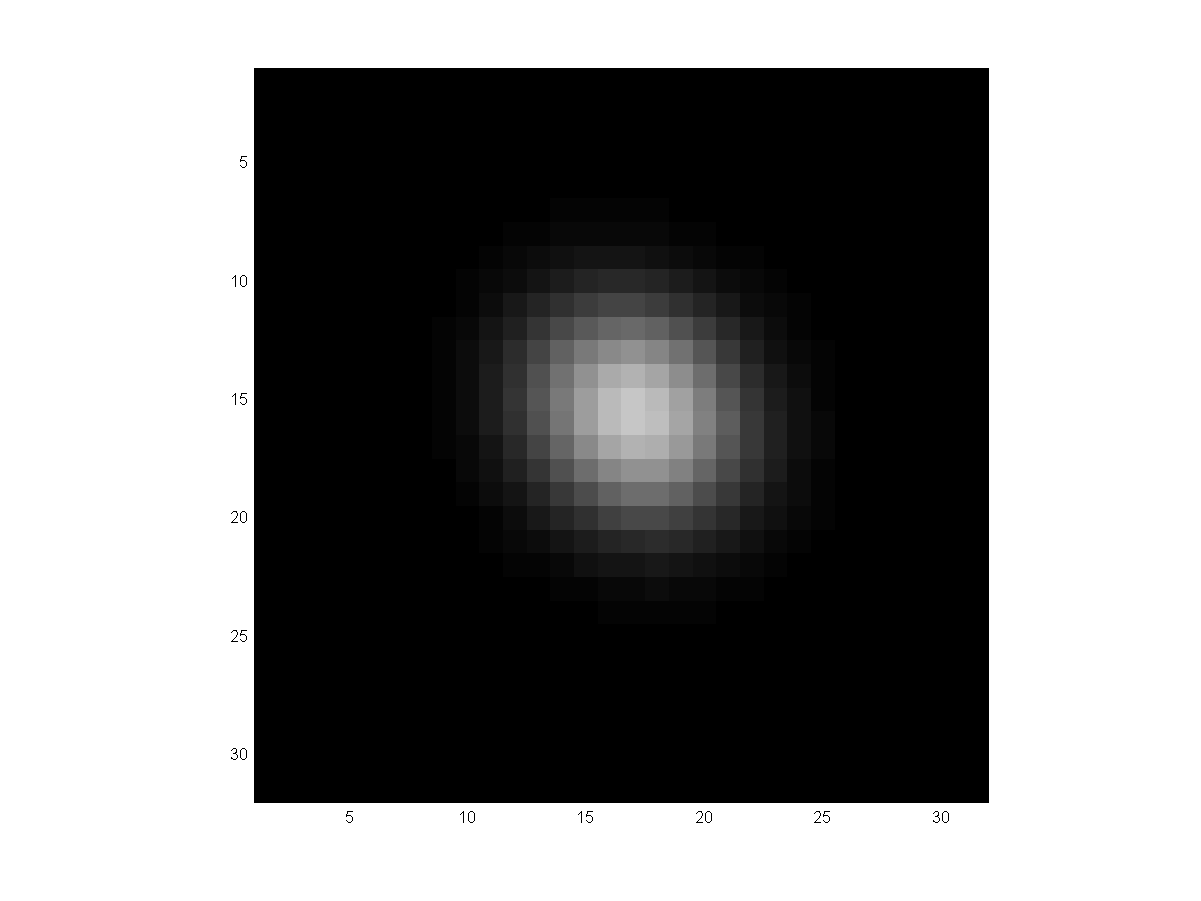
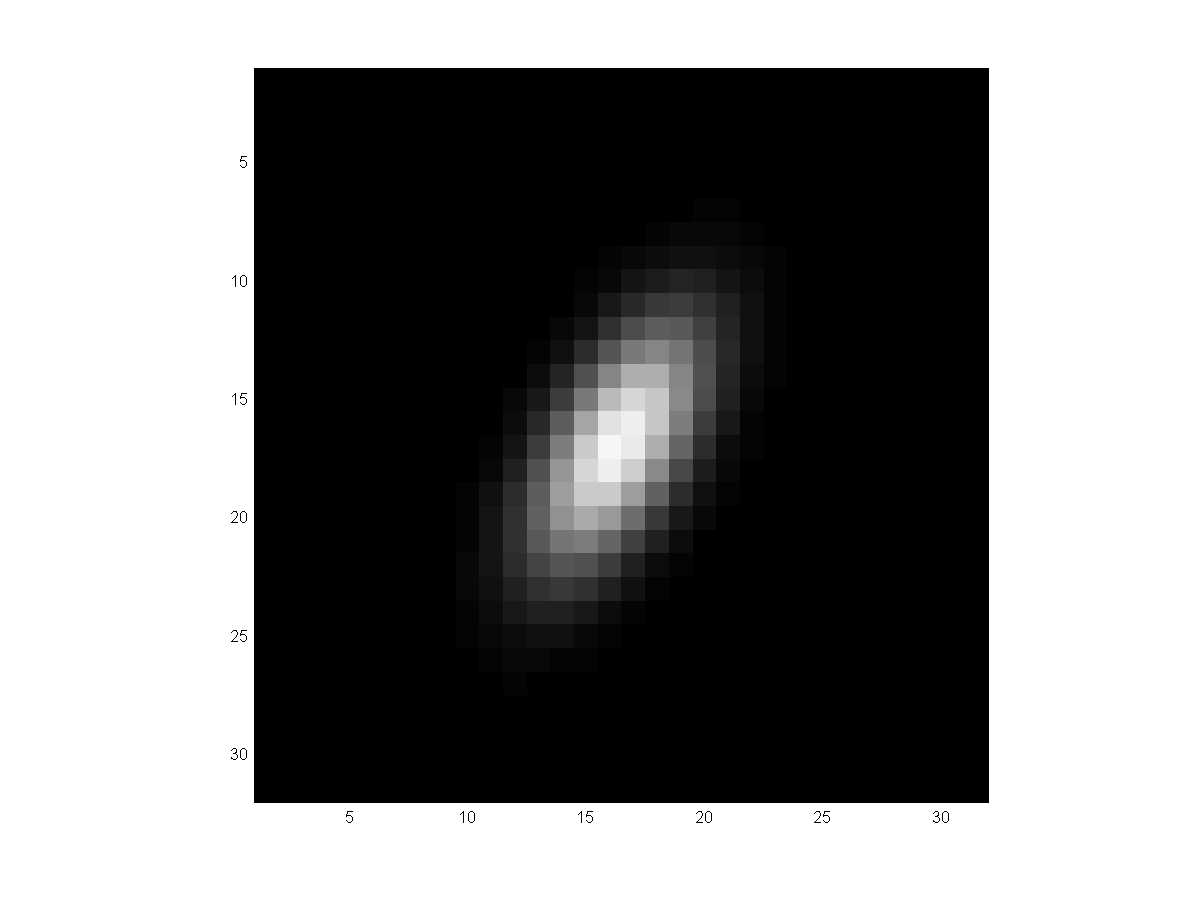
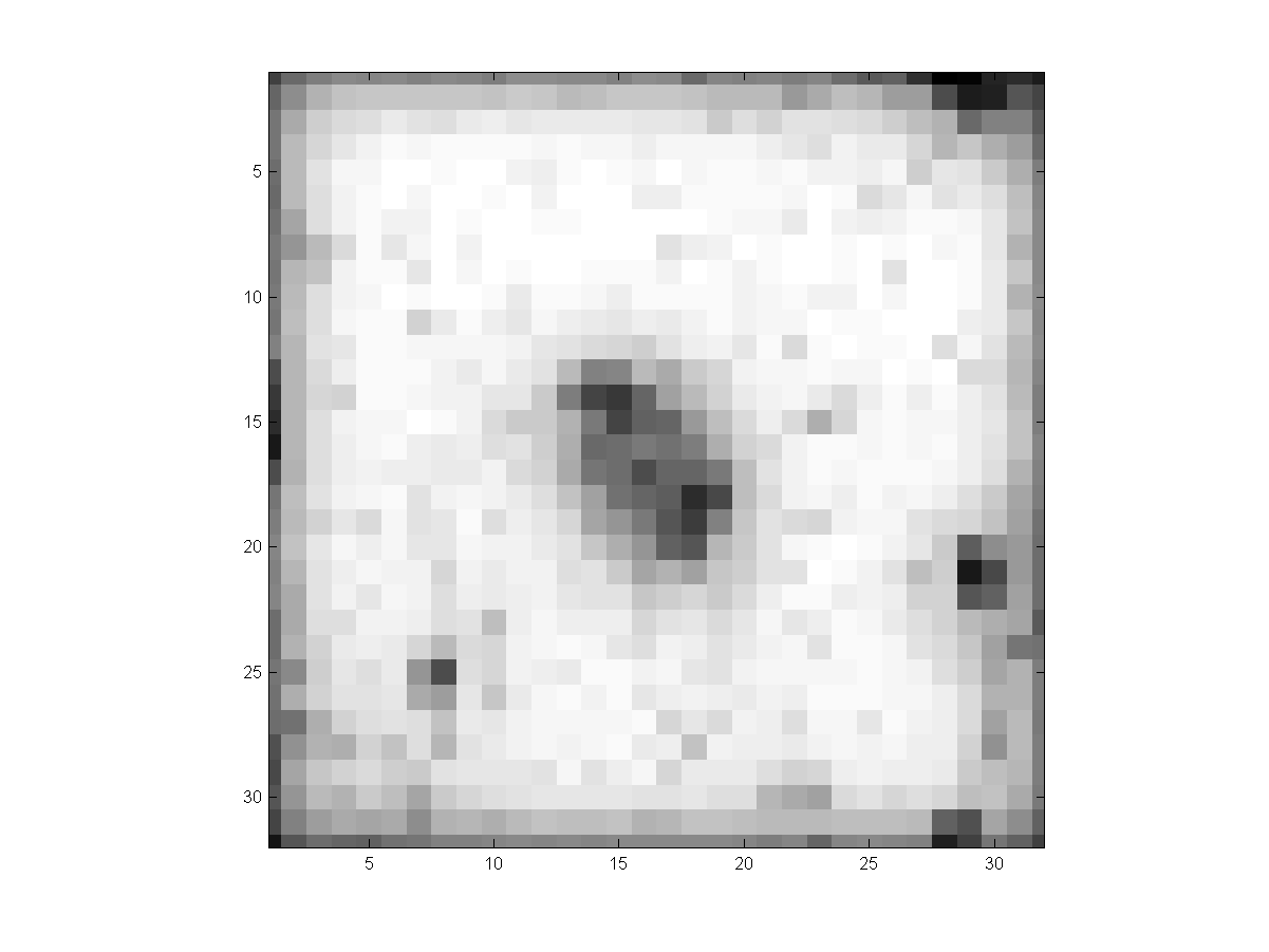
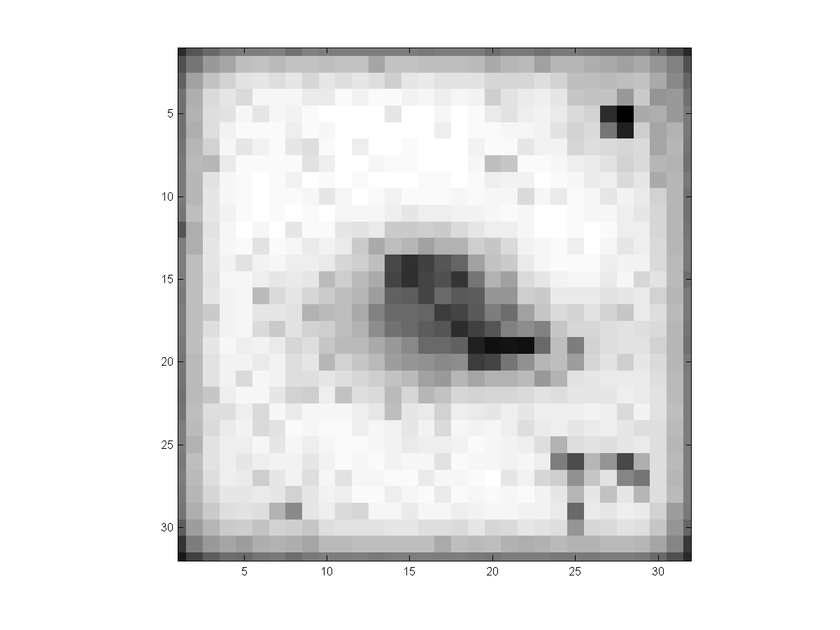
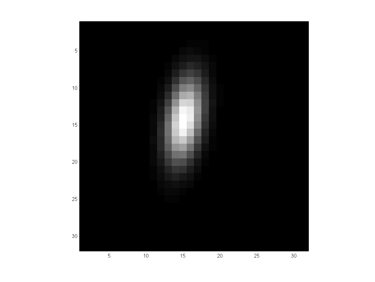
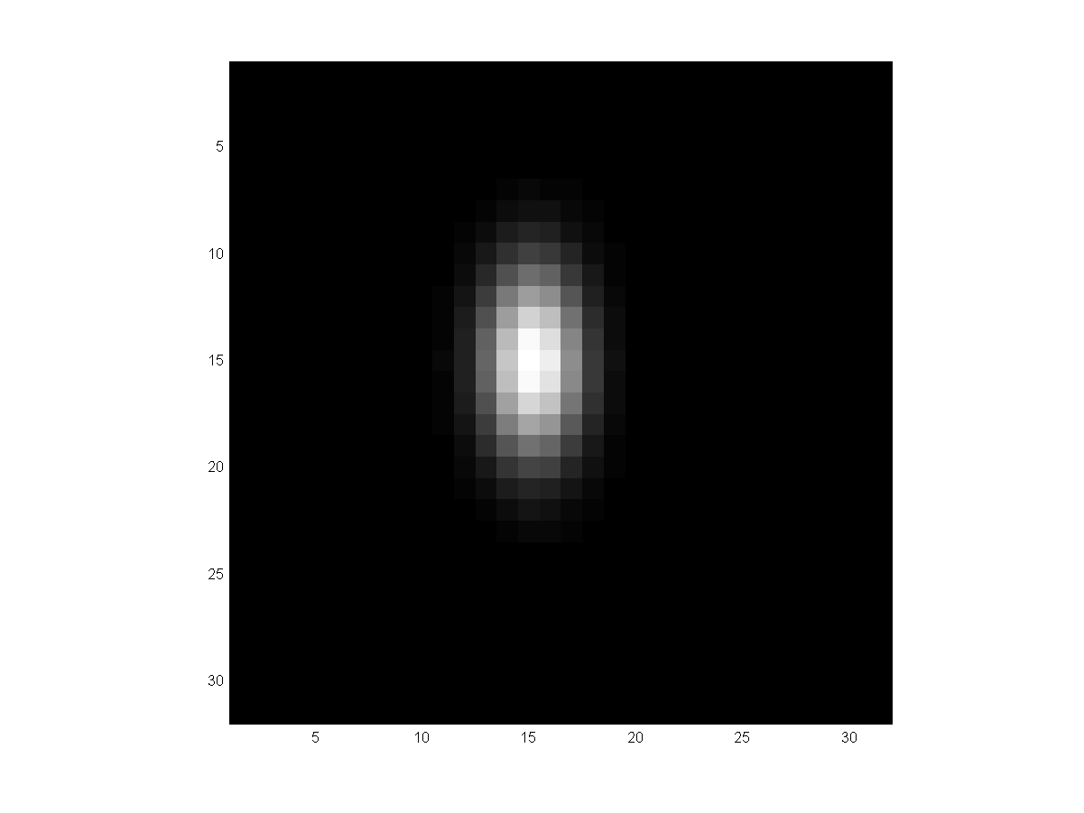
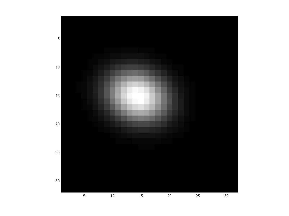
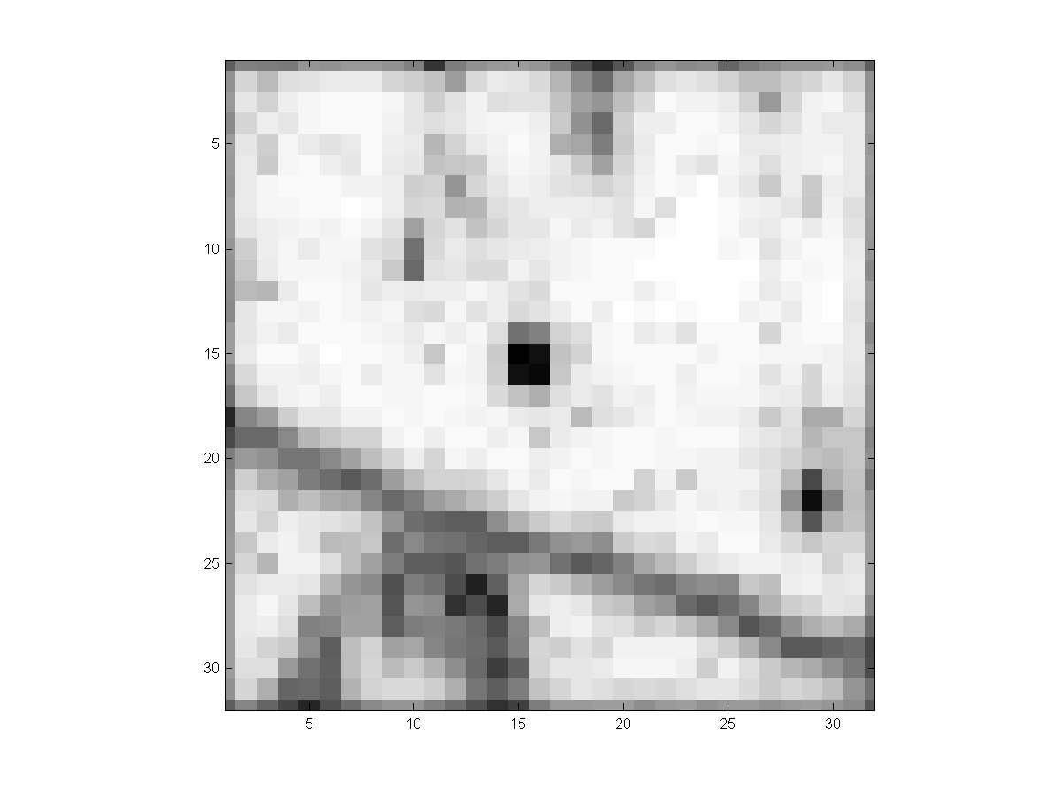
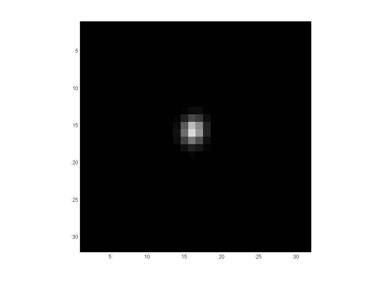
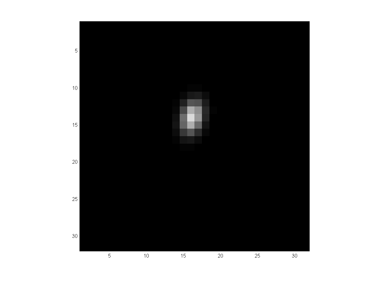
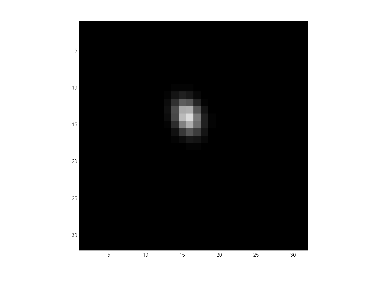
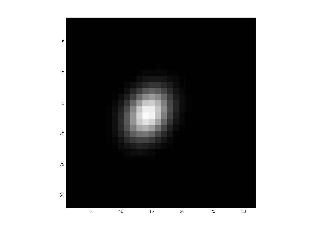
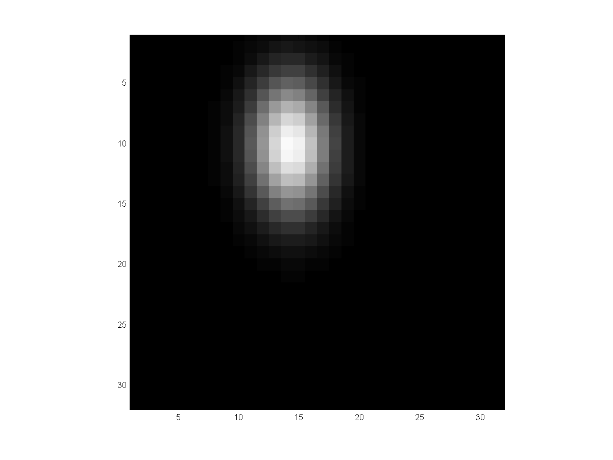
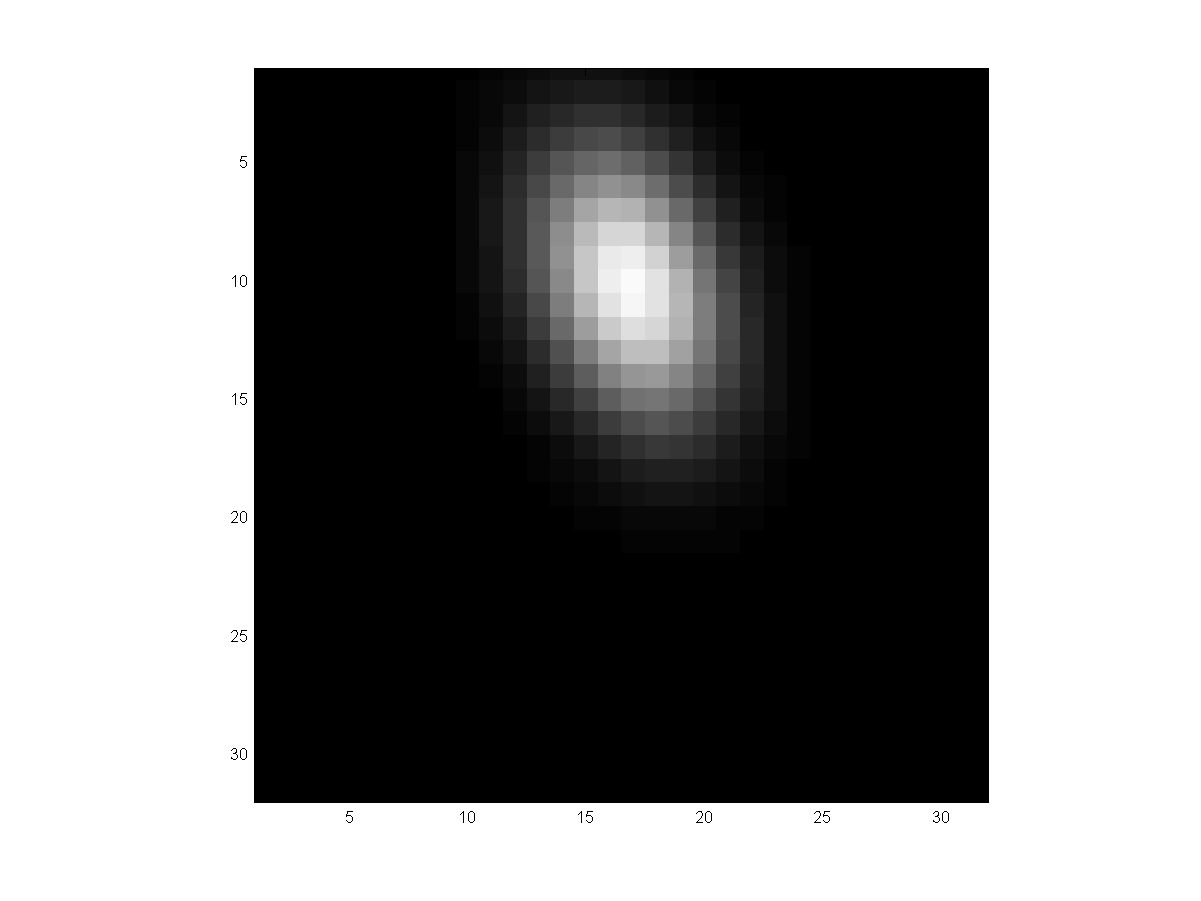
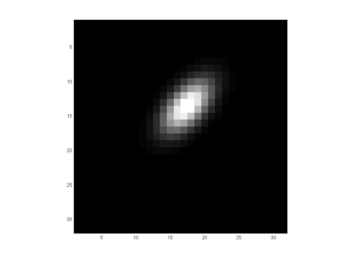
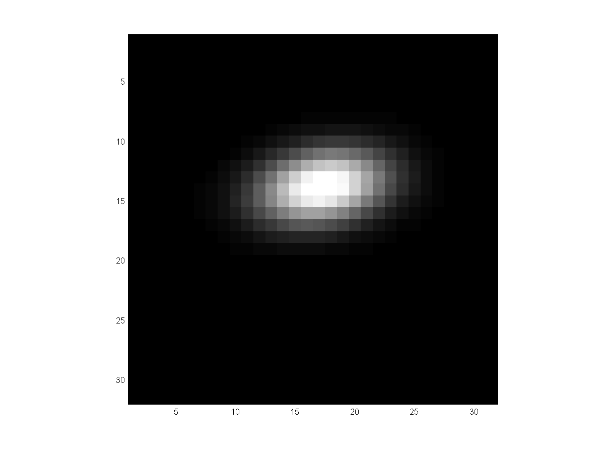
of the segmented tumors. The 3rd columns show the 3D Gaussian density estimated
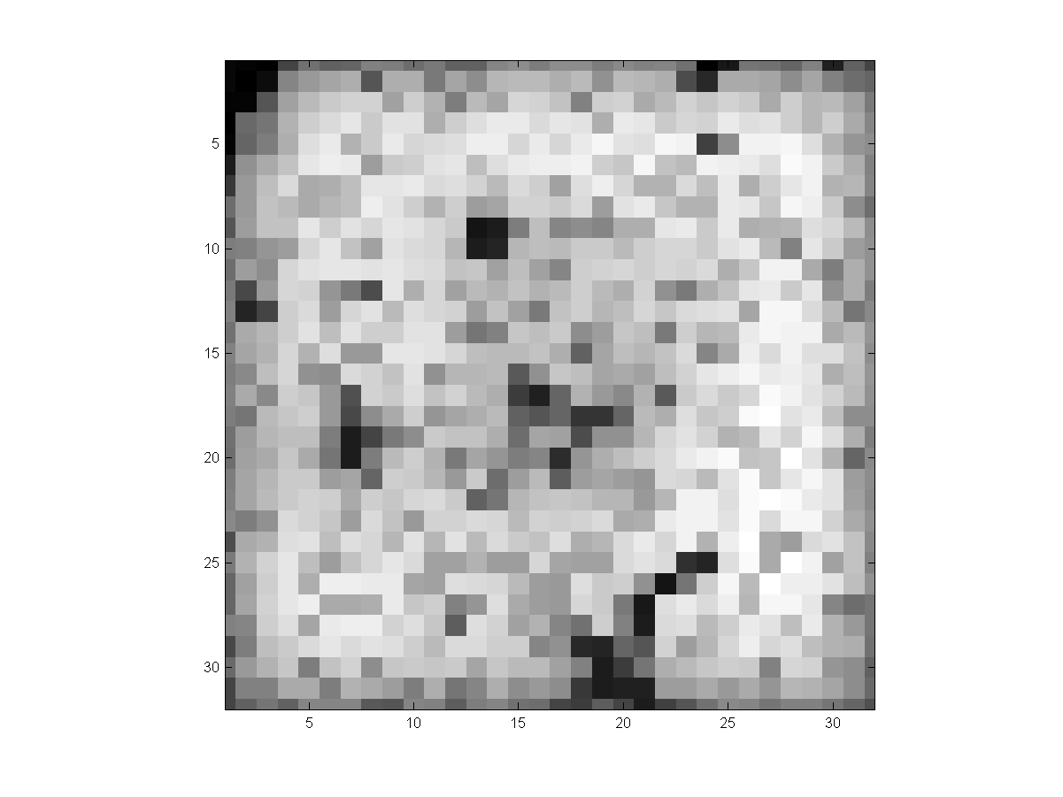
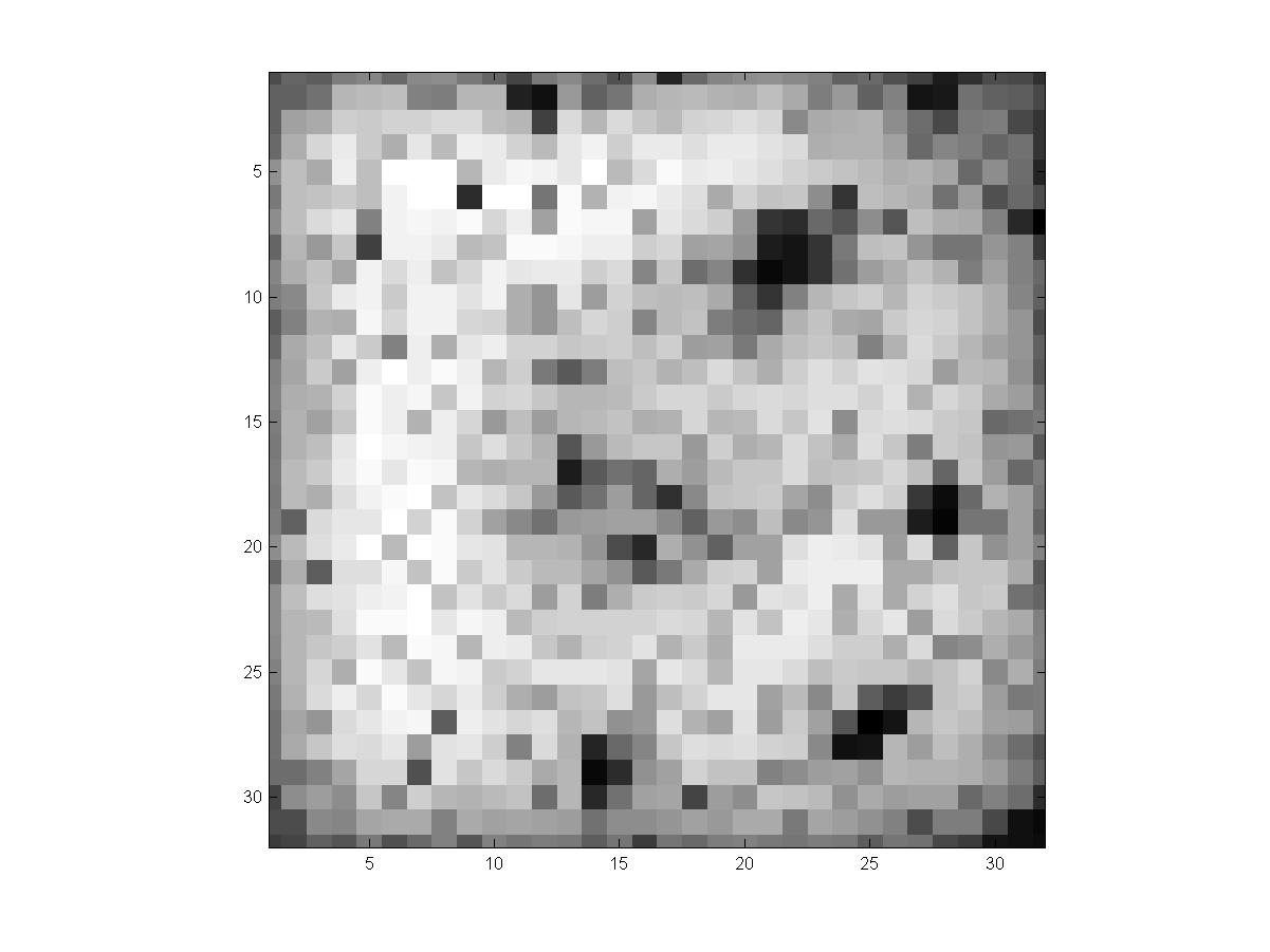
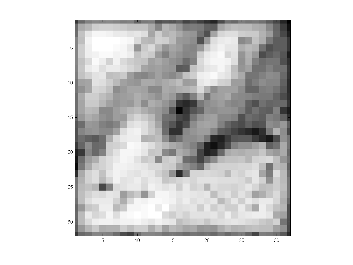
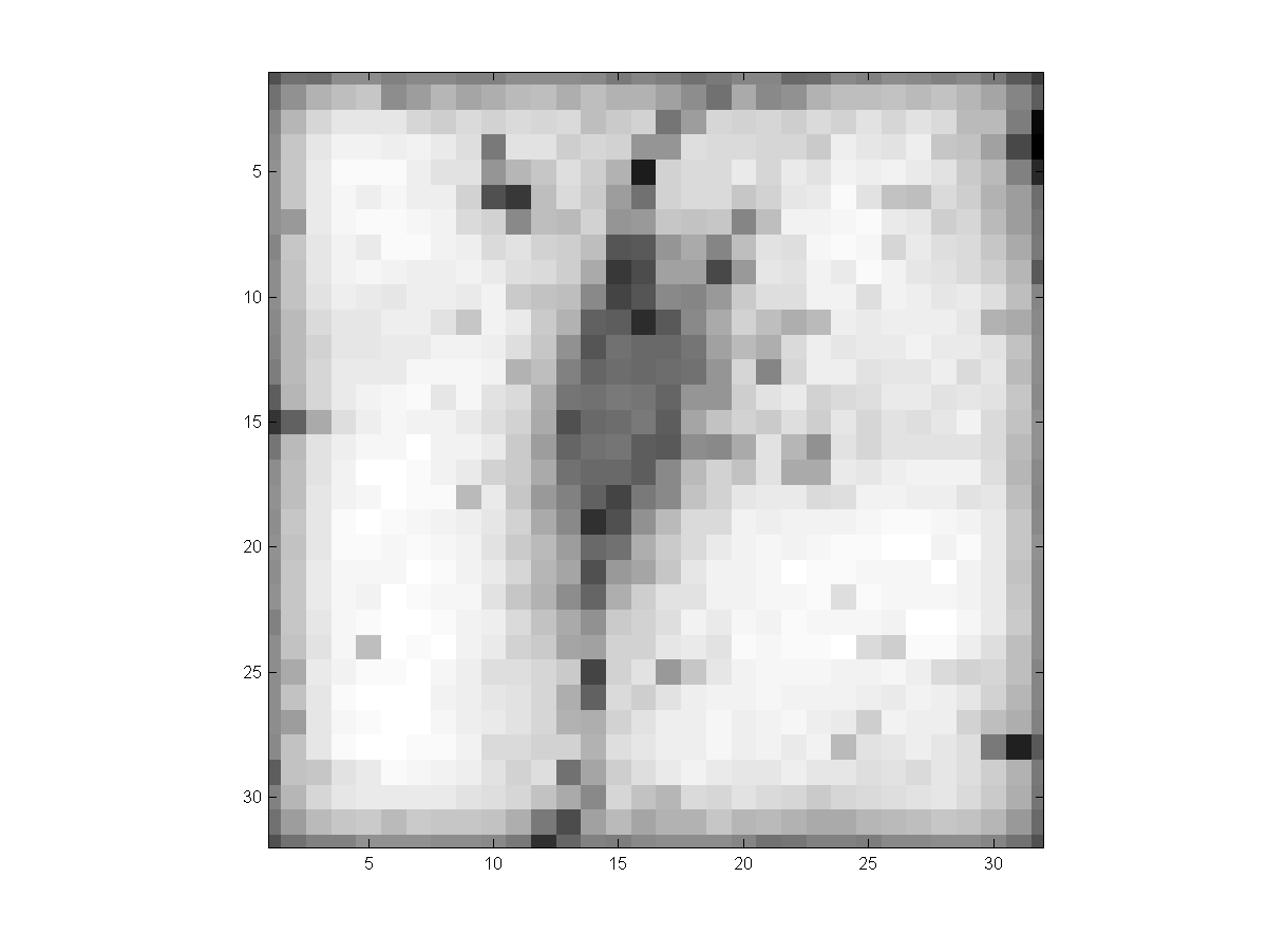
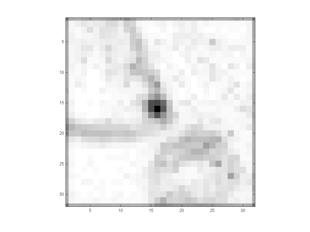
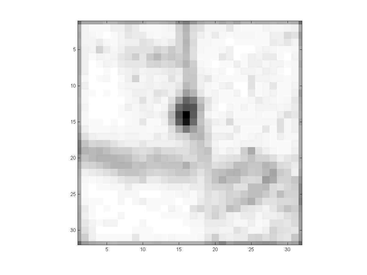
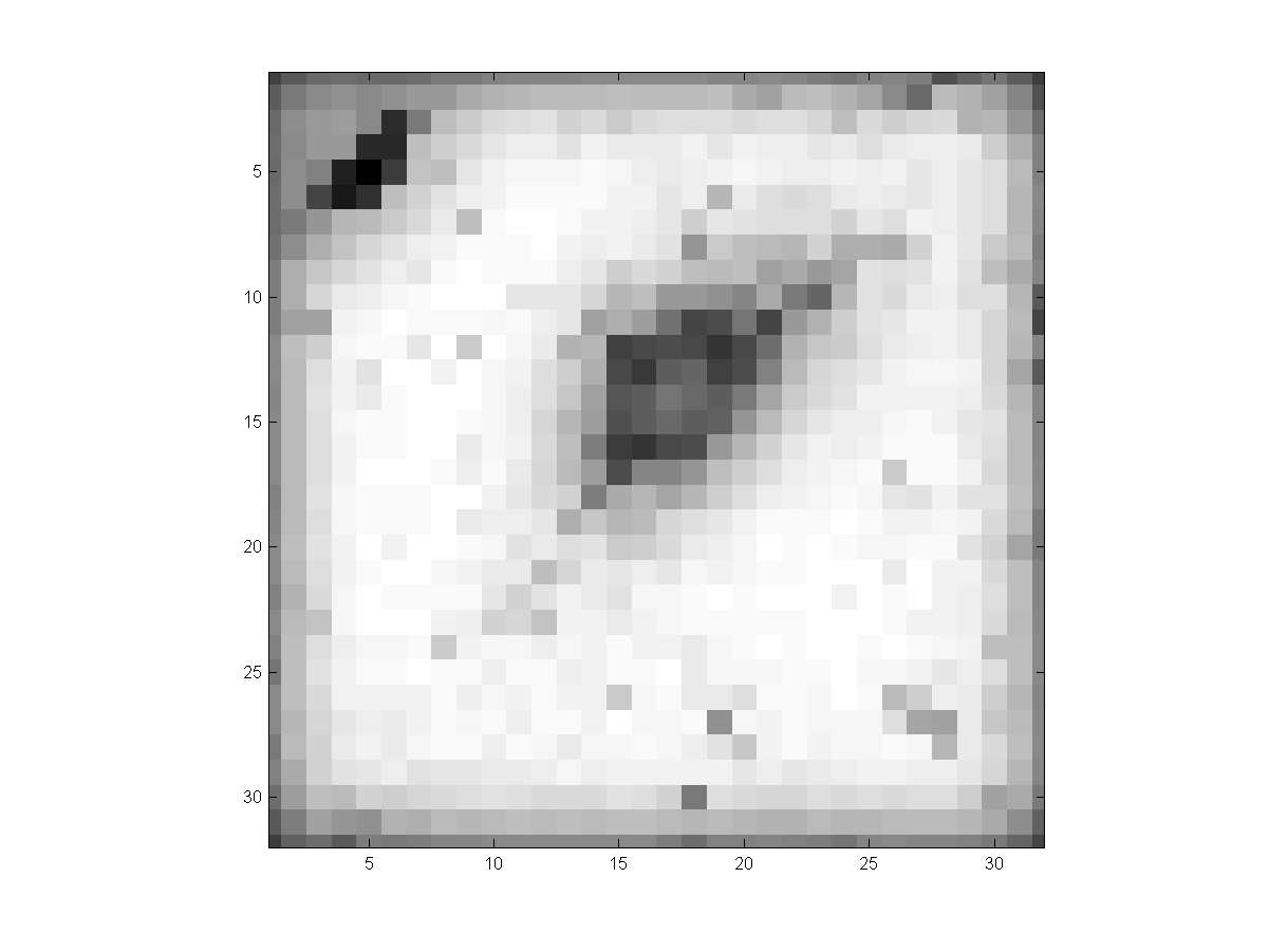
from the first step of our system. The 5th columns show the 3D projection of the
4D joint space density estimates from the second step.
(i) Part- or Non-Solid Nodules
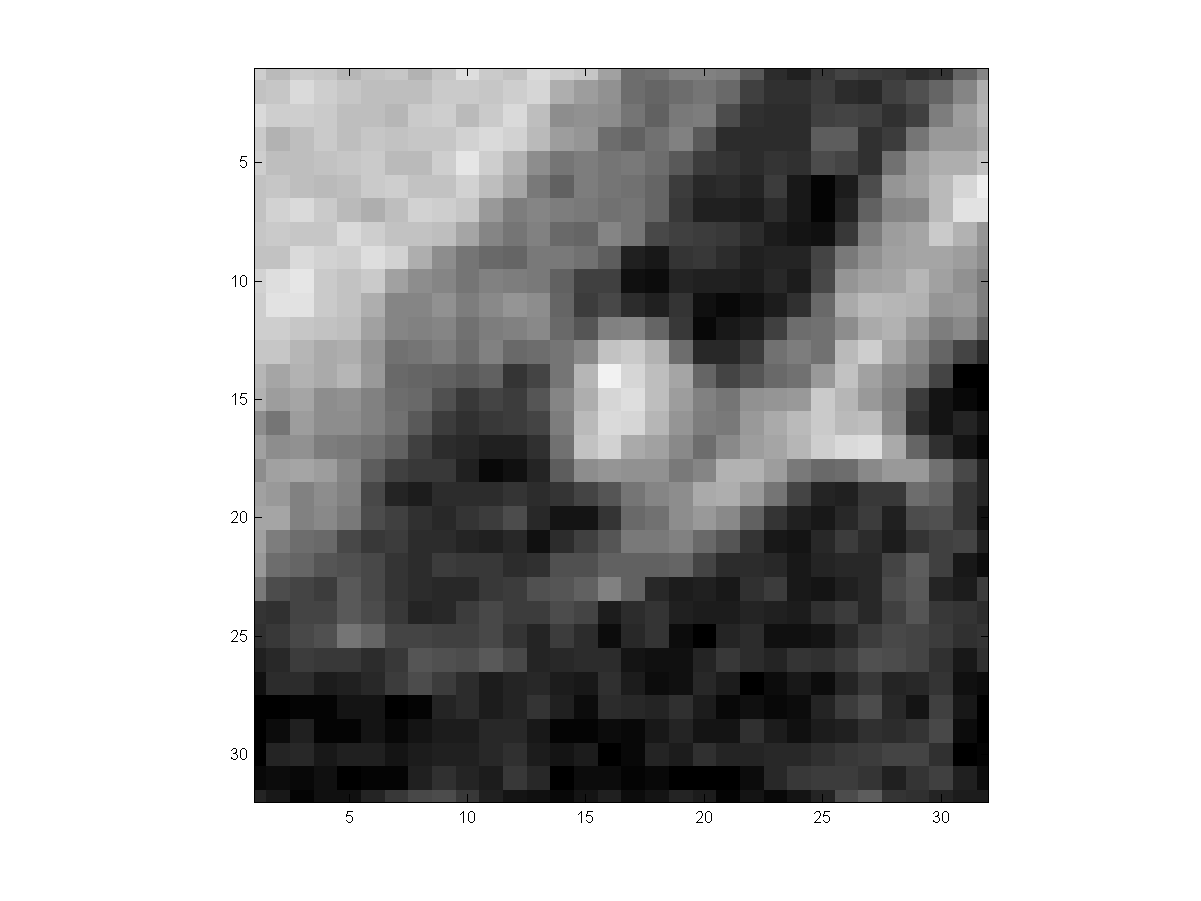
|
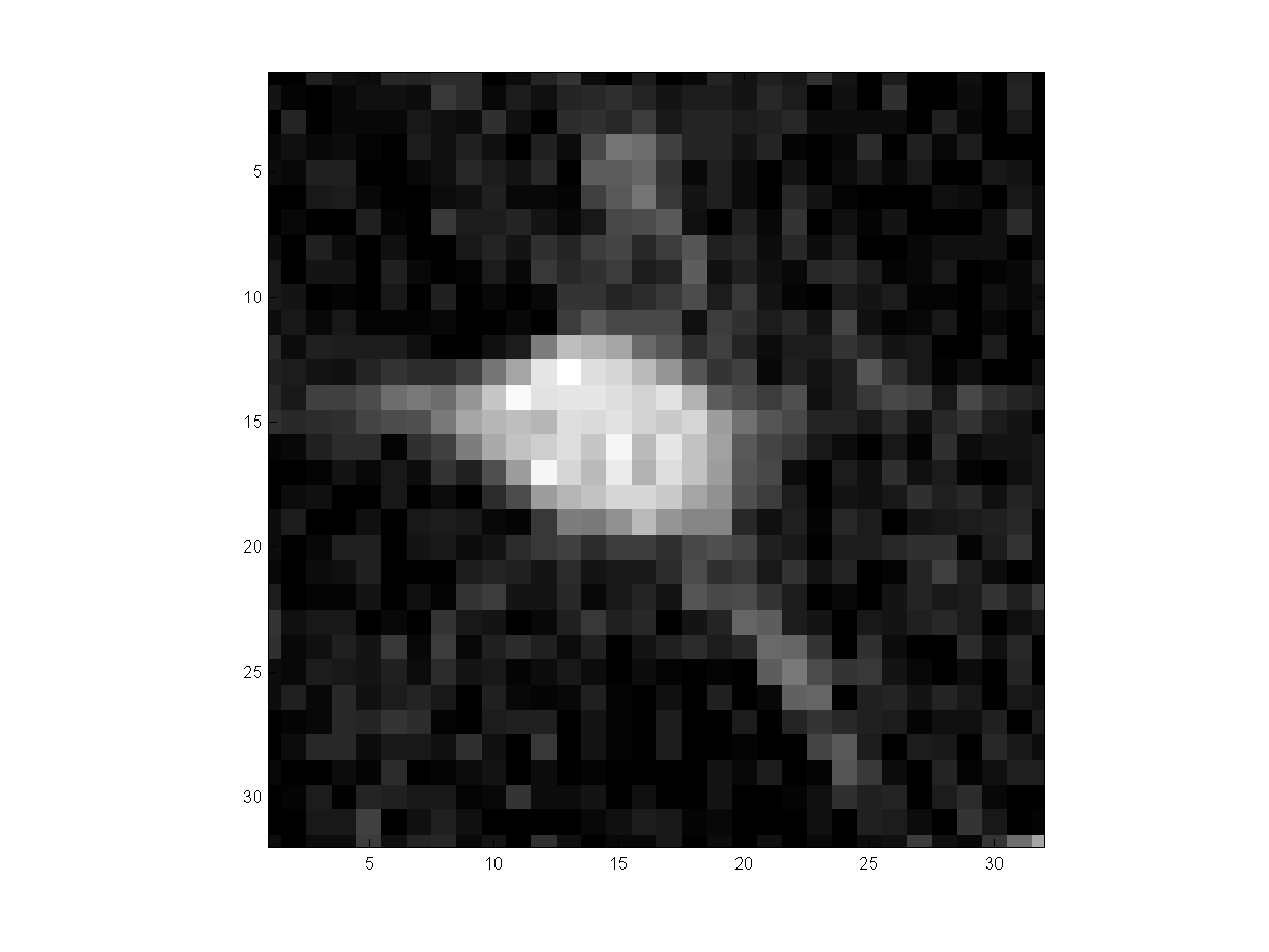
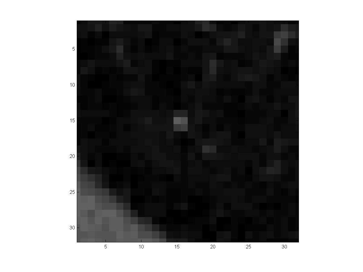
3D Input
|
3D Ellipoidal Fit
|
3D Normal Density Estimate
|
3D Segmentation Result
|
4D Joint Density Estimate
|
yz
|
|
|
|
|
|
xz
|
|
|

|
|
|
xy
|
|
|
|
|
|
|
3D Input
|
3D Ellipoidal Fit
|
3D Normal Density Estimate
|
3D Segmentation Result
|
4D Joint Density Estimate
|
yz
|
|
|
|
|
|
xz
|
|
|
|
|
|
xy
|

|
|
|
|
|
(ii) Pleural Attached
Nodules
|
3D Input
|
3D Ellipoidal Fit
|
3D Normal Density Estimate
|
3D Segmentation Result
|
4D Joint Density Estimate
|
yz
|
|
|

|
|
|
xz
|
|
|
|
|
|
xy
|
|
|
|
|
|
|
3D Input
|
3D Ellipoidal Fit
|
3D Normal Density Estimate
|
3D Segmentation Result
|
4D Joint Density Estimate
|
yz
|
|
|
|
|
|
xz
|
|
|

|
|
|
xy
|
|
|
|
|
|
(iii) Vascularized
Nodules
|
3D Input
|
3D Ellipoidal Fit
|
3D Normal Density Estimate
|
3D Segmentation Result
|
4D Joint Density Estimate
|
yz
|
|
|
|
|
|
xz
|
|
|
|
|
|
xy
|
|
|
|
|
|
|
3D Input
|
3D Ellipoidal Fit
|
3D Normal Density Estimate
|
3D Segmentation Result
|
4D Joint Density Estimate
|
yz
|
|
|
|
|
|
xz
|
|
|
|
|
|
xy
|
|
|
|
|
|
(iv) Small Nodules
|
3D Input
|
3D Ellipoidal Fit
|
3D Normal Density Estimate
|
3D Segmentation Result
|
4D Joint Density Estimate
|
yz
|
|
|

|
|
|
xz
|
|
|

|
|
|
xy
|
|
|
|
|
|
|
3D Input
|
3D Ellipoidal Fit
|
3D Normal Density Estimate
|
3D Segmentation Result
|
4D Joint Density Estimate
|
yz
|
|
|
|
|
|
xz
|

|
|
|
|
|
xy
|
|
|
|
|

|
(v) Examples
|
3D Input
|
3D Ellipoidal Fit
|
3D Normal Density Estimate
|
3D Segmentation Result
|
4D Joint Density Estimate
|
yz
|
|
|
|
|
|
xz
|

|
|
|
|
|
xy
|
|
|
|
|
|
|
3D Input
|
3D Ellipoidal Fit
|
3D Normal Density Estimate
|
3D Segmentation Result
|
4D Joint Density Estimate
|
yz
|
|
|
|
|
|
xz
|
|
|
|
|
|
xy
|
|
|
|
|
|